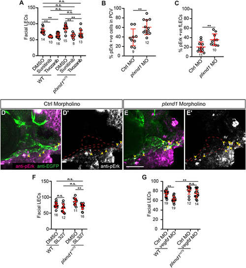
plxnd1 mutants have elevated Vegfr/Erk signalling in the facial lymphatics. (A) Quantitation of facial LEC number in 6 dpf larvae treated from 3 to 6 dpf with either DMSO, 1 µM sunitinib or 2 nM tivozanib. (B,C) Quantitation of the proportion of pErk-positive cells within the PCV at 36 hpf (B) or the facial LECs at 3 dpf (C). (D-E′) Confocal images of anti-pErk (magenta) and anti-EGFP (green) staining in the head at 3 dpf of either control (D,D′) or plxnd1 (E,E′) lyve1b:EGFP morphant larvae. (D′,E′) Anti-pErk staining only. Yellow arrowheads indicate the pErk cells within the facial lymphatics. (F) Quantitation of facial LEC number in 6 dpf larvae treated from 3 to 6 dpf with either DMSO or 5 µM SL327. (G) Quantitation of facial LEC number in lyve1b:DsRed; fli1a:nlsEGFP larvae injected with either control or vegfd morpholinos. P>0.05 (not significant), *P<0.05, **P<0.01 (unpaired t-tests, Kruskal–Wallis and ANOVA); data are mean±s.d. Scale bar: 50 µm. LEC, lymphatic endothelial cell. Numbers in graphs represent numbers of larvae.
|
