Phylogenetic and structural similarity of E piscicida AsdA with other bacteria. (A) Amino acid sequences of AsdA of various bacteria were retrieved from the GenBank database and the unrooted phylogenetic tree of AsdA was constructed by the neighbor-joining method within the MEGAX program. (B) 3D structure of E. piscicida, E. ictaluri and S. Typhimurium AsdA proteins were predicted by using (PS)2: Protein Structure Prediction Server Version 3.0 web portal.

(E) piscicida asdA (χ16000) genotype and phenotype verification. (A) Deletion map of ΔasdA. (B) Genotype verification of E. piscicida ΔasdA (χ16000) by PCR. (C) Phenotypic verification of ΔasdA (χ16000) in the presence or absence of DAP in LB agar plates. (D) Growth curves for J118 and χ16000 in presence or absence of DAP.

Complementation of Δ
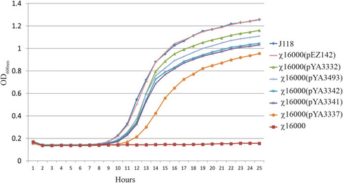
asdA mutant strains with E. piscicida AsdA+ Plasmids. (A)E. piscicida AsdA+ Plasmid maps (pG8R8011-pG8R8018) showing origin of replication, orientation of asdA gene, presence or absence of the promoter for the asdA gene and with either ATG or GTG start codons, Ptrc promoter and multiple cloning sites. (B) Characteristic of E. piscicida AsdA+ plasmids or various strategies to construct AsdA+ plasmids via modification of the plasmid copy number (pUC ori and pBR ori), promoter, SD sequence and start codon. (C) Growth of ΔasdA (χ16000) mutant strains complemented with different E. piscicida AsdA+ plasmids (pG8R8011 - pG8R8018).

Growth of E. piscicida ΔasdA (χ16000) strains complemented with asdA gene from E. ictaluri and Salmonella. Growth of E. piscicida ΔasdA (χ16000) strains complemented with Salmonella AsdA+ plasmids with different copy numbers (high, medium and low) and modified asdA gene promoter.

IAG52B sequence analysis. (A) Alignment of nucleotide and amino acid sequences of the original and codon-optimized IAG52B gene. Line marked with Δ is the codon optimized sequence and * is the original IAG52B sequence. Changed nucleotide sequences are highlighted with blue. (B) Pie chart and table illustrates the amino acid composition of IAG52B. (C) The secondary structures of IAG52B protein were predicted by Protean software by using Gramier-Robson and Chou-Fasman methods. Lines 1, 3, 5, and 7 are the Gramier -Robson methods; the red represents the alpha helix, the green represents the beta fold, the blue represents the turn, the yellow represents the random coil. Lines 2, 4, 6 are for the Chou -Fasman method; the red represents the alpha helix, the green represents the beta fold, the blue for the turn, without random coil prediction. (D) The 3D structure of IAG52B protein was predicted using the Phyre2 web portal.

(A) Vector map of pYA3493 (pBR ori), which is a periplasmic secretion AsdA+ vector. A DNA fragment encoding the β-lactamase signal sequence (bla SS) and 12 amino acid residues of the N-terminus of mature-lactamase was positioned under the control of the Ptrc promoter. Map of pYA3493 shows the unique restriction enzyme sites in the multiple cloning site. (B) Vector map of recombinant plasmid pG8R8029. Codon-optimized gene of IAG52B non-cytoplasmic domain of 1269 bp was PCR amplified and cloned into the pYA3493 at EcoRI-BamHI sites. The IAG52B gene was fused into the same reading frame of bla SS and under the control of the Ptrc promoter. (C) Synthesis of IAG52B antigen in E. piscicida, E. ictaluri and E. coli asdA mutant strains. pYA3493 (vector control) or pG8R8029 (encoding IAG52B) were electroporated into E. piscicida (χ16000) or E. ictaluri (J111) or E. coli (χ6212). IAG52B antigen synthesis was analyzed in these cells by western blotting using an anti-IAG52B antibody. (D) Subcellular location of synthesized IAG52B in E. piscicida and E. coli. Western blot showing IAG52B synthesis in whole cell lysate, periplasmic, cytoplasmic, outer membrane and supernatant fraction of χ16000 and χ6212 harboring pG8R8029. (E) Analysis of IAG52B antigen synthesis in E. piscicida vaccine strain χ16022. pYA3493 (vector control) or pG8R8029 (encoding IAG52B) was electroporated into χ16022, IAG52B antigen synthesis was analyzed by western blotting by using anti-IAG52B antibody.

Dissemination and colonization of χ16022(pG8R8029) in zebrafish tissues. (A) Zebrafish were vaccinated with χ16022(pG8R8029) by i.c. injection. Kidney and spleen were collected from immunized fish at 3- and 5-days post-vaccination (five fish in each group). The tissues were homogenized in 200 μL of BSG and plated on LB agar plates supplemented with 0.2% agarose and 10 μg/mL of colistin. The plates were incubated at 30°C for 48 h and the colonies were counted. The data was a combination of three independent assays.

tnf-α, il-1β, il–6 and il-8 gene expression in χ16022(pG8R8029) vaccinated and control zebrafish. Zebrafish were vaccinated with χ16022(pG8R8029) by i.c. injection. At three, five and seven days of post-immunization, total RNA was extracted from the gills, kidney, intestine and spleen and cDNA was prepared. The qRT-PCR assay was conducted to analyze the expression of tnf-α, il-1β, il-6 and il-8 genes using β-actin as an internal control. The results are expressed as mean ± standard error (bars) from three separate experiments. Differences between uninfected (control) and infected groups were analyzed by two-way ANOVA, where asterisks (*) indicate significant difference (*P < 0.05, **P < 0.01, ****P < 0.0001) with respect to the control group.

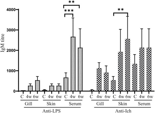
Anti-LPS and anti-Ich antibody responses in zebrafish. Serum and mucosal immunoglobulin M (IgM) responses to E. piscicida lipopolysaccharide (LPS) and Ich IAG52B membrane protein were measured by ELISA at 4 weeks (4w) post-vaccination with χ16022(pG8R8029) and 2 weeks after J118 challenge (6w). The results are expressed as mean ± standard error (bars) from three separate experiments. Differences between treated and control groups were analyzed by two-way ANOVA, where asterisks (*) indicate significant difference (**P < 0.01, ***P < 0.001).

Survival of χ16022(pG8R8029), χ16022(pYA3493) vaccinated and BSG control fish after wild-type E. piscicida challenge. Control and 4 weeks post-vaccination zebrafish were i.c. challenged with 1 × 10⁵ CFU/dose (10 × LD50) of wild-type E. piscicida. Mortality was recorded daily and represented as percent survival.

Acknowledgments
This image is the copyrighted work of the attributed author or publisher, and ZFIN has permission only to display this image to its users. Additional permissions should be obtained from the applicable author or publisher of the image. Full text @ Front Immunol