FIGURE SUMMARY
Title

Loss of zebrafish atp6v1e1b, encoding a subunit of vacuolar ATPase, recapitulates human ARCL type 2C syndrome and identifies multiple pathobiological signatures

Authors
Pottie, L., Van Gool, W., Vanhooydonck, M., Hanisch, F.G., Goeminne, G., Rajkovic, A., Coucke, P., Sips, P., Callewaert, B.
Source
Full text @ PLoS Genet.

<italic toggle='yes'>Atp6v1e1b</italic>-deficient zebrafish have craniofacial, vascular and cardiac abnormalities reminiscent of the human <italic toggle='yes'>ATP6V1E1</italic>-related CL phenotype.

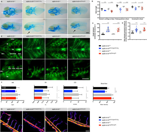
Representative images are shown. (A) Ventral (top panel) and lateral views (bottom panel) of Alcian blue stained craniofacial structures at 5 dpf reveal misshapen and shorter Meckel’s cartilage (m), shorter palatoquadrate (pq) and shorter ceratohyal (ch) structure and a higher angle between ch structures in atp6v1e1b-deficient zebrafish. Pericardial edema is indicated by an arrow. (B) The length of the individual cartilage structures was measured and normalized to the head length of the larvae. (C) Quantification of the angle between the ceratohyal structures. (D) Ratio of the aortic diameter proximal to the bulbus arteriosus (BA) to the average aortic diameter between the first (AA3) and the second branchial arch (AA4) as a measure of local dilatation. (E) Ventral (top panel) and lateral views (bottom panel) of the cardiovascular structure of atp6v1e1b-deficient zebrafish in the Tg(kdrl:eGFP) background at 5 dpf. (F) Cardiac parameter analysis based on brightfield microscopy recordings at 3 dpf. (G) Pseudo-colored processed images representing relative blood flow intensity in the trunk of 3 dpf zebrafish. Major blood vessel ROI used for quantification is marked with a green outline. Data are expressed as mean ± standard deviation (SD). AA1: mandibular arch; AA3: first branchial arch; AA4: second branchial arch; AA5: third branchial arch; AA6: fourth branchial arch; Ao: aorta; BA: bulbus arteriosus; cb1-4: ceratobranchial pairs 1 to 4; ch: ceratohyal; CO: cardiac output; dlav: dorsal longitudinal anastomotic vessel; et: ethmoid plate; h: hyosymplectic; HR: heart rate; isv: intersegmental vessel; m: Meckel’s cartilage; ORA: opercular artery; pcv: posterior caudal vein; pq, palatoquadrate; SV: stroke volume; VA: ventral aorta. Scale bar = 200 μm (A), scale bar = 100 μm (E and G).

Profound epidermal and N-glycosylation alterations in <italic toggle='yes'>atp6v1e1b</italic>-deficient zebrafish.

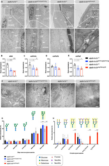
(A) Representative images of ultrathin sections taken from the dermis of atp6v1e1b-deficient zebrafish and WT controls at 4 dpf. Note the two-layered epidermis that is separated from the collagenous stroma of the dermis by a well-defined basement membrane (bm), indicated as dotted line, at this developmental timepoint. In atp6v1e1b-deficient larvae, we observed a larger and more disorganized collagenous stroma of the dermis (atp6v1e1bhi577aTg/hi577aTg) or folded bm (atp6v1e1bcmg78/cmg78). Reproducible results were obtained in three independent experiments. Scale bar = 1 μm (top panel), scale bar = 200 nm (bottom panel). Bm: basement membrane; col: collagenous fibrils; ep: epidermis; m: muscle cell surface; mit: mitochondria; n: nucleus; stroma: primary dermal stroma; rer: rough endoplasmic reticulum. (B-E) Quantification of elnb, col1a1a, col1a1b, col1a2 by RT-qPCR of atp6v1e1b-deficient larvae at 4 dpf compared to WT controls. Data are expressed as mean ± SD from 5 biological replicates. (F) Representative images demonstrating dilated Golgi apparatus in atp6v1e1bcmg78/cmg78. Reproducible results were obtained in three independent experiments. Scale bar = 200 nm (G) Quantification of relative abundancies of MALDI-TOF-MS signals for oligomannose N-glycan species detected in deyolked lysates in atp6v1e1b-deficient zebrafish compared to WT controls. (H) Quantification of relative abundancies of MALDI-TOF-MS signals for complex N-glycan species detected in deyolked lysates in atp6v1e1b-deficient zebrafish compared to WT controls. Data are expressed as mean ± SD from 3 biological replicates. 2-way ANOVA with Tukey test for multiple comparison was used for statistical analysis. Yellow shading indicates differences between WT controls and atp6v1e1b-deficient larvae. Symbols represent monosaccharide residues. Graphical representation is based on the accepted convention from the Symbol Nomenclature for Glycans (Consortium for Functional Glycomics). Yellow circle: Gal; green circle: Man; red triangle: Neu5Ac; light blue diamond: Neu5Gc; Neu5Ac: N-acetylneuraminic acid; Neu5Gc: N-glycolylneuraminic acid.

Effect of loss of <italic toggle='yes'>atp6v1e1b</italic> on the non-canonical function of v-ATPases.

(A) Phosphorylated ribosomal protein S6 kinase 1 (S6K1) (upper panel) and total S6K1 (lower panel) were detected in protein lysates of whole atp6v1e1b-deficient zebrafish and their corresponding WT controls at 4 dpf by immunoblotting. Asterisks indicate non-related, cross-reactive bands. Representative immunoblots are shown. We confirmed equal loading by staining for α-tubulin. (B-D-F) Band intensities of chemiluminescent signals of non-phosphorylated proteins were quantified with ImageJ and normalized to WT controls. (C-E-G) Band intensities of chemiluminescent signals of phosphorylated proteins were quantified with ImageJ. The ratio of phospho-p85-S6K to total p85-S6K, phospho-p70-S6K to total p70-S6K, and phospho-p31-S6K to total p31-S6K was normalized to WT controls. Data are expressed as mean ± SD from 3 biological replicates in B-G. (H-P) Gene expression of Notch-target genes (her4, her6, and hey2), Wnt-target genes (axin2, tcf7, and lef1), and AMPK-target genes (prkaa1, ptch1, and gli1) were investigated by RT-qPCR. Data are expressed as mean ± SD from 5 biological replicates in H-P.

Transcriptomic and metabolomic signature of <italic toggle='yes'>atp6v1e1b</italic> deficiency <italic toggle='yes'>in vivo</italic>.

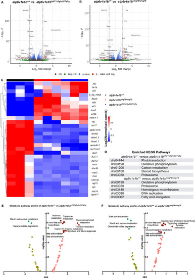
(A) Volcano plot of RNA-sequencing data of atp6v1e1bhi577aTg/hi577aTg. (B) Volcano plot of RNA-sequencing data of atp6v1e1bcmg78/cmg78. The data represents overlapping genes from both ZFIN and Ensembl reference genome mapping (GRCz11). Red spots represent differentially expressed genes (DEGs). The horizontal line denotes the significance threshold (adjusted p<0.05) for DEGs. The vertical line denotes the Log2 fold change (Fc) threshold of 2. (C) Hierarchical clustering of the top 30 most DEGs from whole-body samples of atp6v1e1b-deficient and WT control zebrafish at 3 dpf. DEGs are annotated with gene names of which the protein function is known. DEGs with unknown gene names are listed in S1 Table. Colors range from red (high expression) to blue (low expression). (D) Results of the KEGG pathway analysis showing the most enriched pathways in the differentially expressed gene list of the whole-body samples from the atp6v1e1b-deficient and WT control zebrafish dataset. (E-F) Plots of an integrated analysis based on Metaboanalyst software (pathway tool) for a simplified view of contributing pathways in whole-body samples of atp6v1e1b-deficient zebrafish at 3 dpf. Full analysis based on GSEA parameters and Mummichog parameters are shown in S2 Table. Dark green and brown symbols represent significantly enriched pathways. ND: not determined; NES: normalized enrichment score.

A<italic toggle='yes'>tp6v1e1b</italic> depletion leads to higher amounts of sphingolipids and phospholipids in zebrafish larvae.

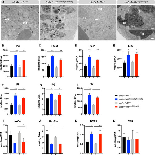
(A) Representative images of ultrathin sections of the yolk from 4 dpf WT control and atp6v1e1b-deficient zebrafish. Atp6v1e1b-deficient larvae reveal an accumulation of electron-dense vesicular bodies. Higher magnification of the lipid whorls is shown in the top right corner of the corresponding image. Scale bar = 500 nm (low magnification), scale bar = 200 nm (high magnification). Results are representative of three independent experiments. Mit: mitochondria; endo: endosomal derived multilamellar bodies; lip: lipid whorls. (B-L) HILIC LC-MS/MS lipidomic analysis of atp6v1e1b-deficient zebrafish and WT controls at 3 dpf. Data are expressed as mean ± SD from 3 biological replicates. PC: phosphatidylcholine; PC-O: 1-alkyl,2-acylphosphatidylcholine; PC-P: 1- alkenyl,2-acylphosphatidylcholine; LPC: lysophosphatidylcholine; PG: phosphatidylglycerol; PI: phosphatidylinositol; SM: sphingomyelin; CER: ceramides; DCER: dihydroceramides; HexCer: hexosylceramides; LacCer: lactosylceramides.

Early respiratory chain deficits resulting from <italic toggle='yes'>atp6v1e1b</italic> depletion <italic toggle='yes'>in vivo</italic>.

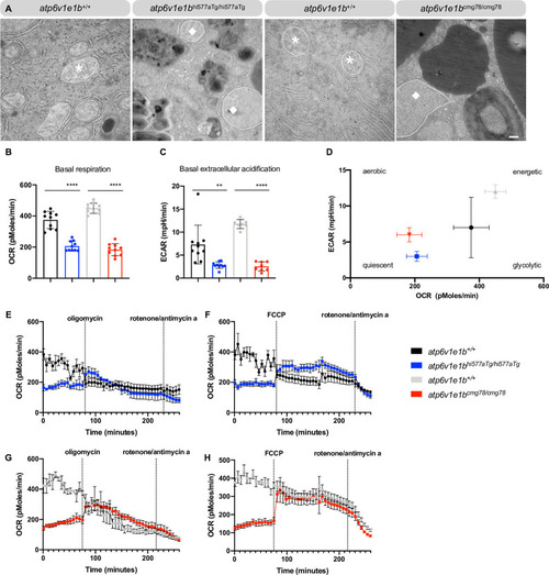
(A) Representative images of ultrathin sections of the yolk from WT control and atp6v1e1b-deficient zebrafish and WT controls at 4 dpf. Asterisk, normal mitochondria. Diamond, dilated mitochondria. Scale bar = 500 nm. Results are representative of three independent experiments. (B) Measurement of the basal respiration and (C) extracellular acidification rate in atp6v1e1bhi577aTg/hi577aTg and atp6v1e1bcmg78/cmg78 zebrafish larvae at 2 dpf compared to WT controls. (D) Plot of OCR vs. ECAR values showed that both mutant zebrafish larvae are in a more quiescent aerobic state than their WTs. (E-H) OCR in WT control and atp6v1e1b-deficient zebrafish after administration of oligomycin and rotenone/antimycin a or FCCP and rotenone/antimycin a. ECAR: extracellular acidification rate; OCR: oxygen consumption rate. Data are expressed as mean ± standard error of the mean (SEM) from 5 biological replicates.

Acknowledgments
This image is the copyrighted work of the attributed author or publisher, and ZFIN has permission only to display this image to its users. Additional permissions should be obtained from the applicable author or publisher of the image. Full text @ PLoS Genet.