IMAGE

Fig 5

ID
ZDB-IMAGE-210714-29
Source
Figures for Pottie et al., 2021
Image
Figure Caption

Fig 5 A<italic toggle='yes'>tp6v1e1b</italic> depletion leads to higher amounts of sphingolipids and phospholipids in zebrafish larvae.

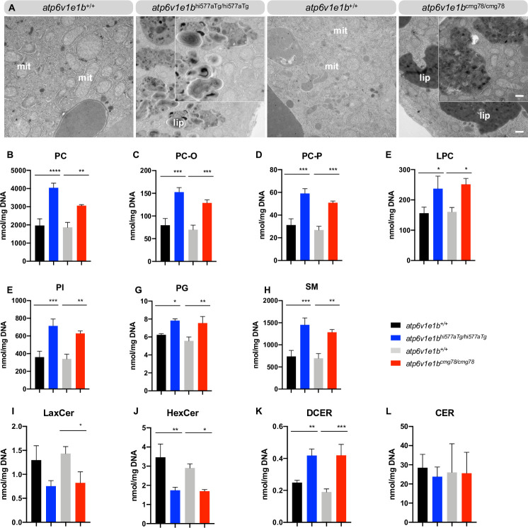
(A) Representative images of ultrathin sections of the yolk from 4 dpf WT control and atp6v1e1b-deficient zebrafish. Atp6v1e1b-deficient larvae reveal an accumulation of electron-dense vesicular bodies. Higher magnification of the lipid whorls is shown in the top right corner of the corresponding image. Scale bar = 500 nm (low magnification), scale bar = 200 nm (high magnification). Results are representative of three independent experiments. Mit: mitochondria; endo: endosomal derived multilamellar bodies; lip: lipid whorls. (B-L) HILIC LC-MS/MS lipidomic analysis of atp6v1e1b-deficient zebrafish and WT controls at 3 dpf. Data are expressed as mean ± SD from 3 biological replicates. PC: phosphatidylcholine; PC-O: 1-alkyl,2-acylphosphatidylcholine; PC-P: 1- alkenyl,2-acylphosphatidylcholine; LPC: lysophosphatidylcholine; PG: phosphatidylglycerol; PI: phosphatidylinositol; SM: sphingomyelin; CER: ceramides; DCER: dihydroceramides; HexCer: hexosylceramides; LacCer: lactosylceramides.

Acknowledgments
This image is the copyrighted work of the attributed author or publisher, and ZFIN has permission only to display this image to its users. Additional permissions should be obtained from the applicable author or publisher of the image. Full text @ PLoS Genet.