FIGURE SUMMARY
Title

Inducible modulation of miR-204 levels in a zebrafish melanoma model

Authors
Sarti, S., De Paolo, R., Ippolito, C., Pucci, A., Pitto, L., Poliseno, L.
Source
Full text @ Biol. Open

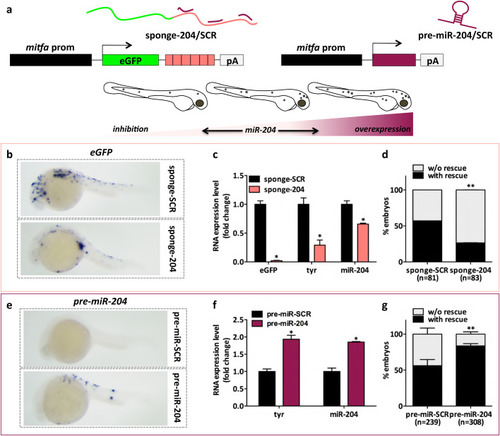
The constitutive modulation of miR-204 expression levels affects melanocyte content in embryos of the Tg(mitfa:BRAFV600E);p53−/−;mitfa−/− line. (A) (upper) Schematic representation of the miniCoopR vectors used for the inhibition and the overexpression of miR-204. On the left, miR-204 inhibition is achieved using a sponge construct that contains six imperfect binding sites for miR-204 downstream of eGFP coding sequence (miniCoopR-sponge-204). On the right, hsa-pre-miR-204 precursor is used to achieve the overexpression of mature miR-204 (miniCoopR-pre-miR-204). As negative controls, miniCoopR-sponge-SCR and miniCoopR-pre-miR-SCR are used, respectively. (lower) Cartoon summarizing the effects observed on embryos’ pigmentation. (B) eGFP mRNA detected by ISH 24 h after the injection of miniCoopR-sponge-SCR or miniCoopR-sponge-204 in 1-cell embryos. (C) eGFP, tyr and mature miR-204 levels measured by qRT-PCR at 7 dpf, upon the injection of miniCoopR-sponge-SCR or miniCoopR-sponge-204 in 1-cell embryos. (D) Percentage of 7dpf embryos showing rescued melanocytes, upon the injection of miniCoopR-sponge-SCR or miniCoopR-sponge-204 at the 1-cell stage. (E) pre-miR-204 RNA detected by ISH 24 h after the injection of miniCoopR-pre-miR-SCR or miniCoopR-pre-miR-204 in 1-cell embryos. (F) tyr and mature miR-204 levels measured by qRT-PCR at 7 dpf, upon the injection of miniCoopR-pre-miR-SCR or miniCoopR-pre-miR-204 in 1-cell embryos. (G) Percentage of 7 dpf embryos showing rescued melanocytes, upon the injection of miniCoopR-pre-miR-SCR or miniCoopR-pre-miR-204 at the 1-cell stage. Statistically significant differences are indicated with asterisks: *P<0.05, **P<0.01.

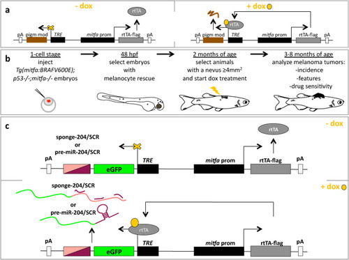
Description of miniCoopR-I vectors for the doxycycline-inducible overexpression of negative and positive modulators of pigmentation. (A) Cartoon describing the general functioning of miniCoopR-I vector. mitfa promoter drives the expression of the reverse tetracycline-controlled transactivator (rtTA, grey). The gene of interest (a pigmentation modulator, brown) is located downstream of tetracycline responsive element (TRE) and gets expressed only when doxycycline (dox, yellow) is added to fish water, so that it can bind to and activate the rtTA. (B) Schematic representation of the experimental protocol to be followed to study the pigmentation modulator of interest. 20–30 pg of miniCoopR-I vector are injected together with 20–30 pg of Tol2 transposase mRNA in 1-cell stage Tg(mitfa:BRAFV600E);p53;mitfa−/− embryos. In line with the protocol described in (Ceol et al., 2011; Iyengar et al., 2012; Painter and Ceol, 2014), successfully injected embryos are selected at 48 hpf on the basis of the presence of rescued melanocytes. Then, at 2 months of age, they are selected on the basis of the presence of nevi ≥4 mm2. At this time point, selected fish are treated with dox (10 uM in fish water), irrespectively of sex. At 3–8 months of age, the indicated parameters are analyzed with the appropriate techniques. (C) Cartoon describing the general functioning of miniCoopR-I-sponge-204/SCR (miR-204 inhibition) and of miniCoopR-pre-miR-204/SCR (miR-204 overexpression).

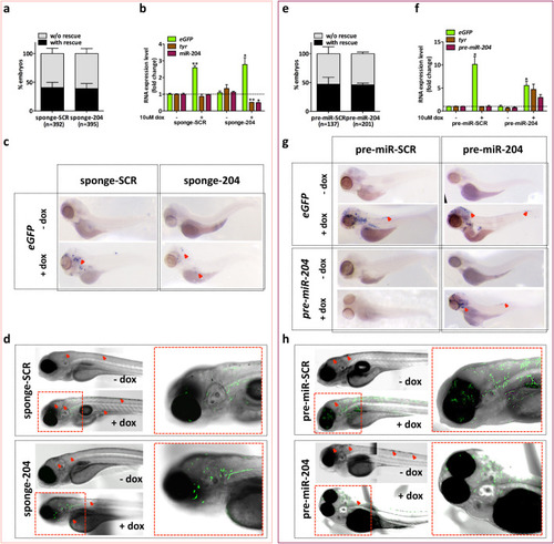
Inducible modulation of miR-204 expression levels in embryos of the Tg(mitfa:BRAFV600E);p53−/−;mitfa−/− line. (A) Embryos injected with miniCoopR-I-sponge-SCR or miniCoopR-I-sponge-204 were observed at 7 dpf. The percentage of embryos with and without melanocyte rescue does not differ significantly between the two experimental groups. (B–D) 1-cell embryos injected with miniCoopR-I-sponge-SCR or miniCoopR-I-sponge-204 were selected at 48 hpf for melanocyte rescue and treated with 10 uM dox for 2 days, then the levels of eGFP, tyr and mature miR-204 were measured by qRT-PCR (B), eGFP mRNA was detected by ISH (C) and eGFP fluorescent protein was detected by confocal microscopy (D). (E) Embryos injected with miniCoopR-I-pre-miR-SCR or miniCoopR-I-pre-miR-204 were observed at 7 dpf. The percentage of embryos with and without melanocyte rescue does not differ significantly between the two experimental groups. (F–H) 1-cell embryos injected with miniCoopR-I-pre-miR-SCR or miniCoopR-I-pre-miR-204 were selected at 48 hpf for melanocyte rescue and treated with 10 uM dox for 2 days, then the levels of eGFP, tyr and pre-miR-204 were measured by qRT-PCR (F), eGFP and pre-miR-204 RNAs were detected by ISH (G) and eGFP fluorescent protein was detected by confocal microscopy (H). Red arrows indicate rescued melanocytes, which are visible independently from dox treatment, * indicates statistically significant differences, *P<0.05, **P<0.01.

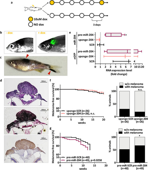
Effect of miR-204 modulation on melanoma incidence. (A–D) Characterization of melanoma tumors that develop in adult fish of the Tg(mitfa:BRAFV600E);p53-/-;mitfa-/- line, injected with miniCoopR-I vectors at 1-cell stage. (A) Schematic representation of the dox-treatment schedule used. (B) eGFP fluorescence is detectable only upon treatment with dox. eGFP fluorescence was detectable in two out of three fish tested, one of which is shown in the pictures. (C) Macroscopic appearance of a fish with melanomas (red arrows). (D) Microscopic features of a melanoma tumor that developed on the head and was excised 1 month after it became visible: H&E (upper), Fontana Masson (middle), anti-BRAFV600E (lower). Images were acquired with a 4x objective. Scale bar: 500um. (E) qRT-PCR quantification of mature miR-204 levels. 1 month after they became visible, tumors that developed on the tail were excised from n=3 fish injected with SCR vectors, n=3 fish injected with miniCoopR-I-sponge-204 vector and n=6 fish injected with miniCoopR-I-pre-miR-204 vector. In all these tumors, effective induction of the transgenic construct was confirmed as higher eGFP mRNA levels compared to a piece of tail that was collected from a non-induced fish and taken as baseline. (F) Melanoma-free survival curve (left) and percentage of animals with/without melanoma (right) upon injection of miniCooR-I-sponge-SCR and miniCoopR-I-sponge-204. (G) Melanoma-free survival curve (left) and percentage of animals with/without melanoma (right) upon injection of miniCooR-I-pre-miR-SCR and miniCoopR-I-pre-miR-204. In melanoma-free survival curves, week count started when the first pulse of 10 uM dox was administered, i.e. at 2 months of age. Statistically significant differences are indicated with asterisks: *P<0.05.

Acknowledgments
This image is the copyrighted work of the attributed author or publisher, and ZFIN has permission only to display this image to its users. Additional permissions should be obtained from the applicable author or publisher of the image. Full text @ Biol. Open