
- Title
-
More Bone with Less Minerals? The Effects of Dietary Phosphorus on the Post-Cranial Skeleton in Zebrafish
- Authors
- Cotti, S., Huysseune, A., Koppe, W., Rücklin, M., Marone, F., Wölfel, E.M., Fiedler, I.A.K., Busse, B., Forlino, A., Witten, P.E.
- Source
- Full text @ Int. J. Mol. Sci.
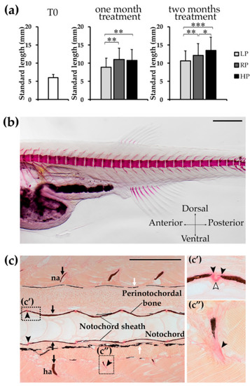
Zebrafish growth and T0 animals. ( |
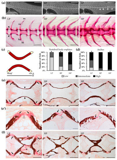
Mineralisation levels of vertebral column after two months of dietary treatment. ( |

Mineralisation levels of median fin structures after two months of dietary treatment. Pterygiophores or radials, endoskeletal structures supporting the dorsal ( |

High dietary P is associated with higher stiffness in the bone formed after two months of treatment. ( |
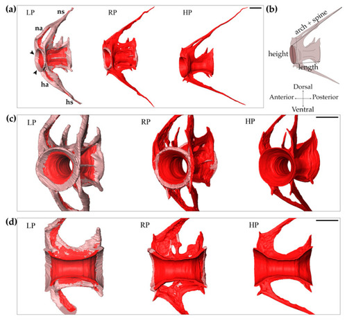
Increased bone formation in LP zebrafish after two months of dietary treatment. Synchrotron X-ray tomographic microscopy scans reveal an increased amount of non-mineralised matrix in the vertebral body and arches of low P diet (LP) treated animals compared to controls (RP) and high P diet (HP) treated individuals. ( |
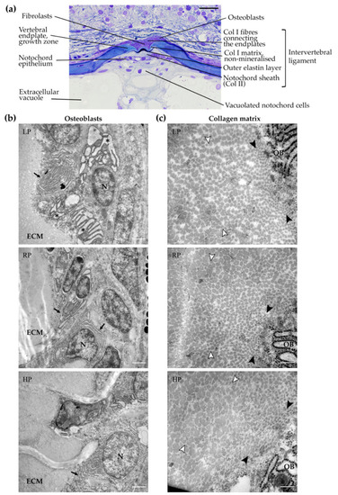
Osteoblasts and collagen type I in the vertebral body endplate growth zone. ( |
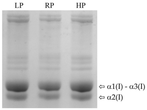
Electrophoretic analysis of collagen type I. Coomassie stained SDS-Urea-PAGE of collagen type I extracted from bone of low P diet (LP) treated zebrafish, controls (RP) and high P diet (HP) treated animals (pool of 2 samples per dietary group). Zebrafish present three collagen type I α chains (α(I)), named α1(I), α3(I) and α2(I). Collagen α(I) chains show bands with similar electrophoretic migration in all dietary groups. |