
- Title
-
The role of endothelial cilia in post-embryonic vascular development
- Authors
- Elworthy, S., Savage, A.M., Wilkinson, R.N., Malicki, J.J., Chico, T.J.A.
- Source
- Full text @ Dev. Dyn.

The Tg(ubi:loxP‐ift54‐loxP‐myr‐mcherry,myl7:EGFP)sh488 rescue transgene provides partial rescue of homozygous ift54 tp49 mutants. A: Diagram of the ubi:loxP‐ift54‐loxP‐myr‐mcherry,myl7:EGFP transgene. B: Five‐dpf siblings from an ift54 tp49/+;Tg(ubi:loxP‐ift54‐loxP‐myr‐mcherry,myl7:EGFP)sh488 x ift54 tp49/+ cross. Sequencing traces show the ift54 tp49 C‐to‐T mutation genotype (asterisk denotes stop codon). Homozygous mutant ift54 tp49 (ift54−/−) embryos without the transgene display the expected curly‐down phenotype. The Tg(ubi:loxP‐ift54‐loxP‐mcherry,myl7:EGFP)sh488 transgene (ubi:ift54) does not affect normal development compared with WT but rescues the phenotype of some ift54 tp49 homozygotes. C: Proportion of phenotypes of the transgenic (+) and nontransgenic (−) embryos from six ift54tp49/+;Tg(ubi:loxP‐ift54‐loxP‐myr‐mcherry,myl7:EGFP)sh488 /+ x ift54 tp49/+ crosses. Total number of embryos is shown above each column. The extent of rescue varied considerably between clutches. D: Five‐month‐old adult fish alongside sequencing traces showing the ift54 tp49 C‐to‐T mutation genotype. ift54 tp49 mutants with the Tg(ubi:loxP‐ift54‐loxP‐myr‐mcherry,myl7:EGFP)sh488 transgene (ift54‐;ubi:ift54) can survive into adulthood but develop scoliosis. Dpf, days postfertilization; WT, wild‐type |
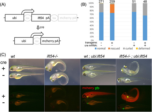
The Tg(ubi:loxP‐ift54‐loxP‐myr‐mcherry,myl7:EGFP)sh488 rescue transgene is recombined after cre mRNA injection, which abolishes its ability to rescue homozygous ift54 tp49 mutants. A: Diagram of the rescue transgene with and without Cre. Cre‐mediated recombination excises WT ift54 and induces mcherry expression. B: Phenotype frequencies in Tg(ubi:loxP‐ift54‐loxP‐myr‐mcherry,myl7:EGFP)sh488 transgenic (+) and nontransgenic (−) embryos from a typical clutch of an ift54 tp49/+;Tg(ubi:loxP‐ift54‐loxP‐myr‐mcherry,myl7:EGFP)sh488 /+ x ift54 tp49/+ cross. Embryos were injected with cre mRNA (+) or uninjected (−). cre injection prevents transgenic rescue. C: Brightfield (top) and fluorescent (bottom) micrographs of 5‐dpf cre mRNA–injected and uninjected embryos from an ift54 tp49/+;Tg(ubi:loxP‐ift54‐loxP‐myr‐mcherry,myl7:EGFP)sh488/+ x ift54 tp49/+ cross. Embryos with the rescue transgene express GFP in the heart. Cre‐mediated recombination prevents rescue of the phenotype and induces mcherry expression. GFP, green fluorescent protein; WT, wild‐type |
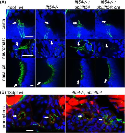
The Tg(ubi:loxP‐ift54‐loxP‐myr‐mcherry,myl7:EGFP)sh488 rescue transgene restores cilia in ift54tp49 mutants. A: Confocal stack projections of the crista of the ear and neuromasts, and confocal slices of the nasal epithelium of 4‐dpf embryos. Acetylated tubulin immunofluorescence of cilia axonemes (green); DAPI staining of DNA in blue. Arrows show cilia axonemes or the positions where they are missing. Images shown are representative of all of the embryos examined of each group from ift54 tp4/+;Tg(ubi:loxP‐ift54‐loxP‐myr‐mcherry,myl7:EGFP)sh488 /+ x ift54 tp49/+ crosses with or without cre mRNA injection. The rescue transgene restores cilia formation in the crista and neuromasts but not nasal epithelium. B: Confocal stack projections of transverse sections of trunk at 13 dpf. Acetylated tubulin (green) immunofluorescence showing cilia axonemes of the pronephric ducts (white arrows), phalloidin staining of actin in red, and DAPI staining of DNA in blue. Images representative of all of three ift54 tp49; Tg(ubi:loxP‐ift54‐loxP‐myr‐mcherry,myl7:EGFP)sh488 fish (ift54−/−;ubi:ift54) and three wt sibs (wt) examined. The rescue transgene rescues cilia of pronephric ducts. WT, wild‐type. Scale bars = 20 μm |
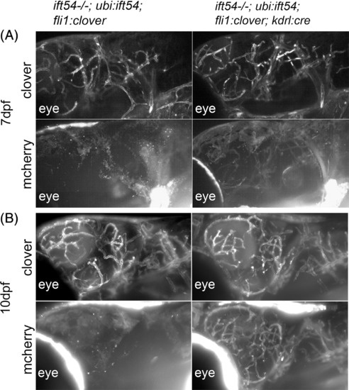
Endothelial‐specific excision of the Tg(ubi:loxP‐ift54‐loxP‐myr‐mcherry,myl7:EGFP)sh488 rescue transgene. A: SPIM stack projections of lateral views of the head of 7‐dfp ift54 tp49; Tg(ubi:loxP‐ift54‐loxP‐myr‐mcherry,myl7:EGFP)sh488 /+;Tg(fli1a:LIFEACT‐clover)sh467 /+ (ift54−/−; ubi:ift54; fli1:clover) and ift54 tp49; Tg(ubi:loxP‐ift54‐loxP‐myr‐mcherry,myl7:EGFP)sh488 /+;Tg(kdrl:cre)s898/+;Tg(fli1a:LIFEACT‐clover)sh467 /+ (itf54−/−; ubi:ift54; fli1:clover; kdrl:cre) fish showing endothelial Clover and faint mcherry expression. The position of the eye is labeled for orientation. Endothelial‐specific Cre‐mediated recombination of the Tg(ubi:loxP‐ift54‐loxP‐myr‐mcherry,myl7:EGFP)sh488 transgene is marked by mcherry expression. B: At 10 dpf, mcherry expression is more pronounced than at 7 dpf EXPRESSION / LABELING:
|

Endothelial‐specific ablation of cilia does not perturb trunk or cerebral vascular development by 10 dpf. A: SPIM stack projections showing endothelial Clover expression in lateral views of the head, anterior trunk, posterior trunk, and tails of 10‐dpf Tg(ubi:loxP‐ift54‐loxP‐myr‐mcherry,myl7:EGFP)sh488/+;Tg(fli1a:LIFEACT‐clover)sh467/+ (wt) (n = 9) and ift54tp49; Tg(ubi:loxP‐ift54‐loxP‐myr‐mcherry,myl7:EGFP)sh488/+;Tg(fli1a:LIFEACT‐clover)sh467/+ (ift54−/−;ubi:ift54) (n = 9) and ift54tp49; Tg(ubi:loxP‐ift54‐loxP‐myr‐mcherry,myl7:EGFP)sh488/+;Tg(kdrl:cre)s898/+;Tg(fli1a:LIFEACT‐clover)sh467/+ (ift54−/−;ubi:ift54;kdrl:cre) (n = 4) fish. B: SPIM stack projections showing endothelial EGFP expression in dorsal views of the head of 10‐dpf Tg(ubi:loxP‐ift54‐loxP‐myr‐mcherry,myl7:EGFP)sh488/+;Tg(fli1a:EGFP)y1/+ (wt) (n = 4) and ift54tp49; Tg(ubi:loxP‐ift54‐loxP‐myr‐mcherry,myl7:EGFP)sh488/+; Tg(fli1a:EGFP)y1/+ (ift54−/−;ubi:ift54) (n = 3) and ift54tp49; Tg(ubi:loxP‐ift54‐loxP‐myr‐mcherry,myl7:EGFP)sh488/+;Tg(kdrl:cre)s898/+; Tg(fli1a:EGFP)y1/+ (ift54−/−;ubi:ift54;kdrl:cre) fish (n = 12). EGFP, enhanced green fluorescent protein; WT, wild‐type |

Endothelial‐specific excision of the Tg(ubi:loxP‐ift54‐loxP‐myr‐mcherry,myl7:EGFP)sh488transgene causes no apparent further impairment to secondary caudal fin vasculature in ift54tp49; Tg(ubi:loxP‐ift54‐loxP‐myr‐mcherry,myl7:EGFP)sh488 fish. A: SPIM stack projections of endothelial cells marked with Clover expression in the caudal fin of 10‐dpf and 14‐dpf Tg(ubi:loxP‐ift54‐loxP‐myr‐mcherry,myl7:EGFP)sh488 /+;Tg(kdrl:cre)s898 /+;Tg(fli1a:LIFEACT‐clover)sh467 /+ fish, showing endothelial‐specific Clover and myr‐mcherry expression. B: SPIM stack projections of endothelial cells marked with Clover in the caudal fin of 14‐dpf ift54 tp49; Tg(ubi:loxP‐ift54‐loxP‐myr‐mcherry,myl7:EGFP)sh488 /+;Tg(fli1a:LIFEACT‐clover)sh467 /+ (ift54−/−; ubi:ift54) and ift54 tp49; Tg(ubi:loxP‐ift54‐loxP‐myr‐mcherry,myl7:EGFP)sh488 /+;Tg(kdrl:cre)s898/+;Tg(fli1a:LIFEACT‐clover)sh467 /+ (ift54−/−; ubi:ift54; kdrl:cre). The secondary caudal fin vasculature forms in the absence of endothelial ift54 function. C: SPIM stack projections of endothelial cells marked with Clover in the caudal fin of 13‐dpf ift54 tp49; Tg(ubi:loxP‐ift54‐loxP‐myr‐mcherry,myl7:EGFP)sh488 /+;Tg(fli1a:LIFEACT‐clover)sh467 /+ (ift54−/−; ubi:ift54) and ift54 tp49; Tg(ubi:loxP‐ift54‐loxP‐myr‐mcherry,myl7:EGFP)sh488 /+;Tg(kdrl:cre)s898 /+;Tg(fli1a:LIFEACT‐clover)sh467 /+ (ift54−/−; ubi:ift54; kdrl:cre) fish. The Tg(kdrl:cre)s898 /+ caused no apparent impairment to the developing fin vascular bed in the fish with partial transgenic rescue of ift54 tp49. Chart shows caudal fin vessel bed relative areas measured from SPIM images. Overlay shows median and interquartile range. Two‐tailed Mann‐Whitney test P = 0.7223 EXPRESSION / LABELING:
|

Endothelial cilia are present in the juvenile caudal fin vascular plexus. Immunofluorescence confocal slices of anti‐acetylated tubulin (red)‐ and anti‐GFP (green)‐stained caudal fin vascular plexus of 11‐dpf Tg(fli1a:LIFEACT‐clover)sh467 /+ (wt) (n = 15), 13‐dpf ift54 tp49; Tg(ubi:loxP‐ift54‐loxP‐myr‐mcherry,myl7:EGFP)sh488 /+;Tg(fli1a:LIFEACT‐clover)sh467 /+ (ift54−/−; ubi:ift54) (n = 4), and 13‐dpf ift54 tp49; Tg(ubi:loxP‐ift54‐loxP‐myr‐mcherry,myl7:EGFP)sh488/+;Tg(kdrl:cre)s898 /+;Tg(fli1a:LIFEACT‐clover)sh467 /+ (ift54−/−;ubi:ift54;kdrl:cre) (n = 4). Slices at shallow and deep levels are shown along with orthogonal views resliced through the confocal stack at the level of cilia marked by arrows. Autofluorescent red blood cells (R) are visible in the vessel lumen. GFP, green fluorescent protein; WT, wild‐type. Scale bar = 10 μm EXPRESSION / LABELING:
|

Endothelial‐specific excision of the Tg(ubi:loxP‐ift54‐loxP‐myr‐mcherry,myl7:EGFP)sh488‐rescuing transgene causes no apparent further impairment to adult vasculature in ift54 tp49; Tg(ubi:loxP‐ift54‐loxP‐myr‐mcherry,myl7:EGFP)sh488 fish. A: Five‐month‐old ift54 tp49; Tg(ubi:loxP‐ift54‐loxP‐myr‐mcherry,myl7:EGFP)sh488 /+ (ift54−/−;ubi:ift54) (n = 24) and ift54 tp49; Tg(ubi:loxP‐ift54‐loxP‐myr‐mcherry,myl7:EGFP)sh488 /+; Tg(kdrl:cre)s898 /+ (ift54−/−;ubi:ift54;kdrl:cre) (n = 26) fish. The Tg(kdrl:cre)s898 /+ caused no apparent further impairment to fish with partial transgenic rescue of ift54 tp49. B: Confocal stack projections showing endothelial EGFP expression in flat‐mount views of swim bladder posterior chamber, pectoral fin, and 4 day‐postamputation regenerated caudal fin from sibling four‐month‐old Tg(ubi:loxP‐ift54‐loxP‐myr‐mcherry,myl7:EGFP)sh488 /+;Tg(fli1a:EGFP)y1 /+ (n = 5) (wt) and ift54 tp49; Tg(ubi:loxP‐ift54‐loxP‐myr‐mcherry,myl7:EGFP)sh488 /+; Tg(fli1a:EGFP)y1 /+ (representative of four animals analyzed) (ift54−/−;ubi:ift54) and ift54 tp49; Tg(ubi:loxP‐ift54‐loxP‐myr‐mcherry,myl7:EGFP)sh488/+;Tg(kdrl:cre)s898 /+; Tg(fli1a:EGFP)y1 /+ (n = 5) (ift54−/−;ubi:ift54;kdrl:cre) fish. Yellow lines indicate approximate position of original fin amputation. EGFP, enhanced green fluorescent protein; WT, wild‐type. Scale bars = 300 μm |

Endothelial‐specific excision of the Tg(ubi:loxP‐ift54‐loxP‐myr‐mcherry,myl7:EGFP)sh488‐rescuing transgene causes no apparent further impairment to adult retina vasculature in ift54tp49; Tg(ubi:loxP‐ift54‐loxP‐myr‐mcherry,myl7:EGFP)sh488 fish. Confocal stack projections showing endothelial EGFP expression in flat‐mount views of retina from sibling four‐month‐old Tg(ubi:loxP‐ift54‐loxP‐myr‐mcherry,myl7:EGFP)sh488 /+;Tg(fli1a:EGFP)y1 /+ (n = 5) (wt) and ift54tp49; Tg(ubi:loxP‐ift54‐loxP‐myr‐mcherry,myl7:EGFP)sh488 /+; Tg(fli1a:EGFP)y1 /+ (n = 4) (ift54−/−;ubi:ift54) and ift54 tp49; Tg(ubi:loxP‐ift54‐loxP‐myr‐mcherry,myl7:EGFP)sh488 /+;Tg(kdrl:cre)s898/+; Tg(fli1a:EGFP)y1 /+ (n = 5) (ift54−/−;ubi:ift54;kdrl:cre) fish. The yellow boxes indicate the region of retina enlarged below. The Tg(kdrl:cre)s898 /+ caused no apparent further impairment to the vasculature in the fish with partial transgenic rescue of ift54 tp49. EGFP, enhanced green fluorescent protein; WT, wild‐type. Scale bars = 300 μm |
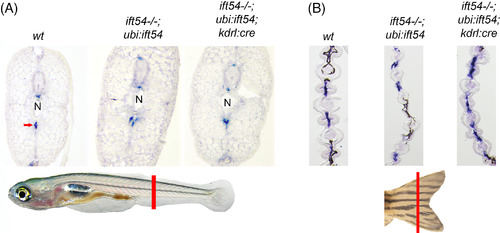
Endothelial‐specific excision of the Tg(ubi:loxP‐ift54‐loxP‐myr‐mcherry,myl7:EGFP)sh488‐rescuing transgene causes no apparent further impairment to tagln‐expressing perivascular mural cells in ift54 tp49; Tg(ubi:loxP‐ift54‐loxP‐myr‐mcherry,myl7:EGFP)sh488 fish. A: In situ hybridization for tagln expression on transverse posterior trunk frozen sections of 13‐dpf wt (wt) and sibling ift54 tp49; Tg(ubi:loxP‐ift54‐loxP‐myr‐mcherry,myl7:EGFP)sh488 /+ (ift54−/−;ubi:ift54) and ift54 tp49; Tg(ubi:loxP‐ift54‐loxP‐myr‐mcherry,myl7:EGFP)sh488 /+; Tg(kdrl:cre)s898/+ (ift54−/−;ubi:ift54;kdrl:cre) fish. The red line in the cartoon illustrates where sections were taken. Tagln expression is detected at the site of arteries such as the dorsal aorta (arrow). The Tg(kdrl:cre)s898 /+ caused no apparent further impairment to perivascular mural cells in the fish with partial transgenic rescue of ift54 tp49. Images representative of four animals analyzed for each genotype. B: In situ hybridization for tagln expression on transverse caudal fin frozen sections of adult wt (wt) and sibling ift54 tp49; Tg(ubi:loxP‐ift54‐loxP‐myr‐mcherry,myl7:EGFP)sh488 /+ (ift54‐;ubi:ift54) and ift54 tp49; Tg(ubi:loxP‐ift54‐loxP‐myr‐mcherry,myl7:EGFP)sh488 /+; Tg(kdrl:cre)s898 /+ (ift54‐;ubi:ift54;kdrl:cre) fish. The red line in the cartoon illustrates where sections were taken. The Tg(kdrl:cre)s898 /+ caused no apparent further impairment to tagln expression in the fish with partial transgenic rescue of ift54 tp49. Images representative of four animals analyzed for each genotype. WT, wild‐type |
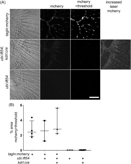
Endothelial‐specific excision of the Tg(ubi:loxP‐ift54‐loxP‐myr‐mcherry,myl7:EGFP)sh488‐rescuing transgene causes no apparent further impairment to tagln‐expressing perivascular mural cells in ift54 tp49; Tg(ubi:loxP‐ift54‐loxP‐myr‐mcherry,myl7:EGFP)sh488 fish expressing a Tg(tagln:mcherry)sh441 transgene. A: Confocal stack projections showing mcherry fluorescence from 14‐dpf caudal fins of Tg(tagln:mcherry)sh441 /+ (tagln:mcherry), Tg(ubi:loxP‐ift54‐loxP‐myr‐mcherry,myl7:EGFP)sh488 /+;Tg(kdrl:cre)s898 (ubi:ift54;kdrl:cre), and the control background fluorescence signal from unrecombined sibling Tg(ubi:loxP‐ift54‐loxP‐myr‐mcherry,myl7:EGFP)sh488 /+ (ubi:ift54) fish. Also shown are binary threshold processed images as used for quantification of the area of Tg(tagln:mcherry)sh441 /+ expression. Single‐focal‐plane transmitted light views are shown for orientation. The microscope settings suitable for Tg(tagln:mcherry)sh441 /+ detect insignificant fluorescence from Tg(ubi:loxP‐ift54‐loxP‐myr‐mcherry,myl7:EGFP)sh488 /+;Tg(kdrl:cre)s898, but endothelial mcherry is detected at increased laser power. B: Chart showing % of field of view with >threshold mcherry expression as measured from processed confocal projections of caudal fins from fish with the different transgenes as shown in representative examples in A. Overlay shows median and interquartile range. Scale bar A = 100 μm EXPRESSION / LABELING:
|

Endothelial‐specific excision of the Tg(ubi:loxP‐ift54‐loxP‐myr‐mcherry,myl7:EGFP)sh488‐rescuing transgene causes no apparent further impairment to perivascular mural cells in the caudal fin vascular plexus of ift54 tp49; Tg(ubi:loxP‐ift54‐loxP‐myr‐mcherry,myl7:EGFP)sh488 fish. A: Confocal stack projections showing mcherry fluorescence from 17‐dpf caudal fins of ift54tp4; Tg(ubi:loxP‐ift54‐loxP‐myr‐mcherry,myl7:EGFP)sh488 /+; Tg(tagln:mcherry)sh441 /+ (ift54−/−; ubi:ift54; tagln:mcherry) and ift54 tp4; Tg(ubi:loxP‐ift54‐loxP‐myr‐mcherry,myl7:EGFP)sh488/+;Tg(kdrl:cre)s898; Tg(tagln:mcherry)sh441 /+ (ift54−/−; ubi:ift54; tagln:mcherry; kdrl:cre). Also shown are binary threshold processed images as used for quantification of the area of Tg(tagln:mcherry)sh441 /+ expression. Single‐focal‐plane transmitted light views are shown for orientation. B: Chart shows % of field of view with >threshold mcherry expression as measured from processed confocal projections of caudal fins. Overlay shows median and interquartile range. Two‐tailed Mann‐Whitney test P = 0.5362. The Tg(kdrl:cre)s898 /+ caused no apparent impairment to mural cell coverage in the newly developed fin vascular bed in the fish with partial transgenic rescue of ift54 tp49 EXPRESSION / LABELING:
|