FIGURE SUMMARY
Title

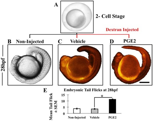
Prostaglandin E2 promotes embryonic vascular development and maturation in zebrafish

Authors
Ugwuagbo, K.C., Maiti, S., Omar, A., Hunter, S., Nault, B., Northam, C., Majumder, M.
Source
Full text @ Biol. Open

Microinjection and embryonic movement at 28 hpf. (A) We collected zebrafish eggs at the two-cell stage and a few non-injected embryos were kept as a reference (B). Only vehicle (C) and PGE2 (D) embryos were fluorescently labeled due to dextran injection. We monitored post-injection growth of embryos with both stereo and fluorescent microscopes and observed an increase in embryonic tail flicks in the PGE2 group (shown in Movie 1A-C). (E) From each biological replicate we selected some embryos to measure tail flicks. Chart showing the mean of embryonic tail flicks of non-injected (n=17), vehicle (n=26) and PGE2 (n=26) injected embryos±s.e.m. We conducted an unpaired t-test and results show a significant increase in tail flicks in PGE2 injected group compared to the vehicle with a two-tailed unpaired t-test, *P=0.0001. Scale bar: 5 μm in A; 25 μm in B,C,D.

PGE2 increases zebrafish embryonic development and pigmentation at 48 hpf. For the measurement of pigmentation, the gray-scaled images from the stereomicroscope were converted to red-colored scale using ImageJ. The red color intensity of the pigmented areas in the vehicle (A) was used as a threshold to measure PGE2-induced pigmentation (B). We selected a few embryos from three biological replicates for this quantification. (C) Data are represented as mean of embryonic pigmentation for PGE2 (n=10) and vehicle (n=11) groups±s.e.m. An unpaired t-test comparing means showed a significant upregulation of embryonic pigmentation in PGE2 group with a two-tailed unpaired t-test, *P=0.0001. (E) The PGE2-treated fish is larger with the tail almost reaching the head in the embryonic sac (dotted red line with arrow) compared to the vehicle (D). We measured the areas of the fish body (denoted with yellow dotted line) and yolk sac (denoted with green dotted line) and calculated the ratio of the areas as body/yolk. (F) The chart shows a very significant growth difference between the PGE2 and vehicle groups with a two-tailed unpaired t-test, **P=0.0086. Scale bar: 10 μm.

PGE2 promotes early hatching of zebrafish at 50 hpf. (A–C) Representative images of hatched embryos in non-injected (A), vehicle (B) and PGE2 injected (C) groups. Scale bar: 10 μm. We analyzed n=90 non-injected, n=61 vehicle and n=90 PGE2 embryos. (D) Data are presented as the mean of percentages (hatched/total number of eggs) of hatched embryos±s.e.m. Hatched embryo numbers were significantly high in PGE2 (33%) compared with the vehicle (5%), with a two-tailed unpaired t-test, **P=0.0001. (E) We also recorded the larval movements as a measure of swimming activity post-hatching in all three groups (shown in Movie 2A-C) at 53 hpf. Data presented as the mean of larval movements of hatched embryos [non-injected (n=14), vehicle (n=31) and PGE2 (n=35)]±s.e.m. We conducted an unpaired t-test showing significantly higher motility in PGE2 group compared to the vehicle with a two-tailed unpaired t-test, **P=0.0008.

PGE2 induced vascular maturation and increased heart rate in zebrafish. (A,B) Gray-scaled images of trunk vasculature of developing zebrafish in both vehicle and PGE2 groups at 96 hpf. Both vehicle and PGE2 groups were microinjected with Texas-Red dextran dye and grown under the same conditions, with vasculature formation captured from 53 hpf to 96 hpf, data presented only for 96 hpf. (C,D) Fluorescence images of trunk vasculature in the vehicle and PGE2 larvae. Vehicle color was considered as a threshold to measure fluorescence of the PGE2 using ImageJ. (E) The chart showing the mean of trunk vascular fluorescence measured for both vehicle (n=9) and PGE2 (n=9) larvae±s.e.m. The PGE2 group showed a significant (*P=0.0001) increase in fluorescence compared to the vehicle group. The PGE2-injected larvae also showed a clear formation of the mature vasculature with dorsal longitudinal anastomosing vessel (DLAV), intersegmental arteries (ISA), dorsal aorta (DA) and posterior cardinal vein (PCV) formation while the vehicle remained premature. (F) The chart shows the mean heart rate of selective embryos from three biological replicates (n=12 for vehicles and n=15 for PGE2)±s.e.m. An unpaired t-test showed a significantly high heart rate in the PGE2 group compared to the vehicle group larvae at 72 hpf, with a two-tailed unpaired t-test, **P=0.0221. Video data for non-injected presented in Movie 3A, vehicle injected in Movie 3B and PGE2 injected in Movie 3C. Scale bar: 10 μm.

PGE2 induces angiogenesis in zebrafish in a time-dependent manner. The dextran dye can trace cell lineage to track vascular development in whole fish. However as the embryo matures, the dye starts to diffuse, which makes it difficult to capture vascular maturation beyond 96 hpf. (A–F) The developing zebrafish trunk vasculature in both vehicle and PGE2 groups from 53 hpf to 96 hpf. The region of interest (ROI) in white dotted boxes shows the difference in vascular fluorescence between groups. (F) In this image, the ROI is expanded to show the dorsal aorta (DA), dorsal longitudinal anastomosing vessel (DLAV), posterior cardinal vein (PCV) and accompanying intersegmental arteries (ISA). (G) We selected a few zebrafish embryos [PGE2 (n=9) and vehicle (n=9)] in both groups to measure fluorescence intensity with ImageJ. The chart represents the mean fluorescence intensity±s.e.m. An unpaired t-test was conducted for each time point and we observed a significant increase in the trunk vasculature fluorescence in the PGE2 compared to the vehicle group at 72 hpf and 96 hpf, respectively. *P=0.007, **P=0.0001. Scale bar: 15 μm.

We captured vascular development of hatched zebrafish embryos at different time points 53, 72 and 96hpf with a fluorescent microscope. A region of interest (ROI) for vascular maturation was shown with white dotted rectangle boxes. Vascular development for the vehicle at three-time points are presented in A, C, E and for PGE2 presented in B, D, F.


Acknowledgments
This image is the copyrighted work of the attributed author or publisher, and ZFIN has permission only to display this image to its users. Additional permissions should be obtained from the applicable author or publisher of the image. Full text @ Biol. Open