
- Title
-
Identification of DEAD-Box RNA Helicase DDX41 as a Trafficking Protein That Involves in Multiple Innate Immune Signaling Pathways in a Zebrafish Model
- Authors
- Ma, J.X., Li, J.Y., Fan, D.D., Feng, W., Lin, A.F., Xiang, L.X., Shao, J.Z.
- Source
- Full text @ Front Immunol

ZFIN is incorporating published figure images and captions as part of an ongoing project. Figures from some publications have not yet been curated, or are not available for display because of copyright restrictions. |
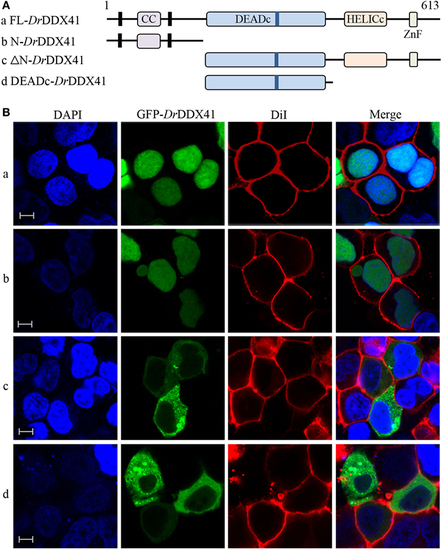
Subcellular localization of Danio rerio DDX41 (DrDDX41) and identification of the nuclear localization signal motifs within it. (A) Schematic diagram of full-length (FL) and DrDDX41 mutants with the coiled-coil (CC), DEADc, HELICc, ZnF_C2HC domains, and residue numbers as indicated. The various DrDDX41 fragments were inserted into the C-terminus of pEGFP-N1. (B) Representative images of transfected HEK293T cells. The N-terminal 190 amino acids targeted GFP to the nucleus (a,b). Deletion of this region in DrDDX41 (191–613, c) and DrDDX41 (191–411, d) resulting in punctate cytoplasmic staining and exclusion from the nucleus. Scale bars represent 5 µm. Images were captured under a laser-scanning confocal microscope (Zeiss LSM-710; original magnification, 630×). |
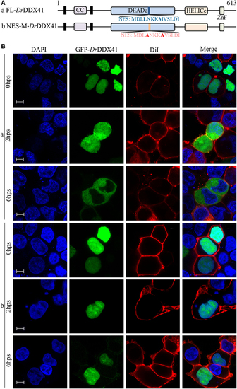
Trafficking analysis of Danio rerio DDX41 (DrDDX41) and identification of the nuclear export signal (NES) motif within it. (A) Schematic diagram of full-length and the NES-mutated DrDDX41. (B) Trafficking of DrDDX41 in response to poly(dA:dT) stimulation at different hours post stimulation (hps) and the contribution of NES motif to the trafficking. Stimulation with poly(dA:dT) induced transportation of DrDDX41 (full-length, a) from nucleus to cytoplasm. Meanwhile, the NES-like-mutant DrDDX41 (NES-M, b) remained in the nucleus without any response to poly(dA:dT) stimulation. Scale bars represent 5 µm. Images were captured under a laser-scanning confocal microscope (Zeiss LSM-710; original magnification, 630×). |
|
ZFIN is incorporating published figure images and captions as part of an ongoing project. Figures from some publications have not yet been curated, or are not available for display because of copyright restrictions. PHENOTYPE:
|
|
ZFIN is incorporating published figure images and captions as part of an ongoing project. Figures from some publications have not yet been curated, or are not available for display because of copyright restrictions. EXPRESSION / LABELING:
|
|
ZFIN is incorporating published figure images and captions as part of an ongoing project. Figures from some publications have not yet been curated, or are not available for display because of copyright restrictions. PHENOTYPE:
|

Subcellular localization analysis of DrDDX41 in zebrafish WBCs and ZF4 cells. (A) Western blot shows that the rabbit anti-DrDDX41 Ab can specifically bind to the corresponding endogenous target proteins in the whole cell lysates of the WBCs sorted from the peritoneal blood, kidney, and spleen. (B) Confocal microscopy image of WBCs stained with rabbit anti-DrDDX41 Ab, showing that the DrDDX41 protein partially distributed in nucleus and cytoplasm in the natural WBCs. WBCs stained with a non-related rabbit IgG was used as negative control. (C) Representative images of transfected ZF4 cells with pEGFP-N1 and pEGFP-DrDDX41. Nuclei were stained with the DNA-intercalating dye DAPI. Scale bars represent 5 μm. Images were captured under a laser scanning confocal microscope (Zeiss LSM-710; original magnification, 630×). EXPRESSION / LABELING:
|