
- Title
-
Inhibitory neuron migration and IPL formation in the developing zebrafish retina
- Authors
- Chow, R.W., Almeida, A.D., Randlett, O., Norden, C., Harris, W.A.
- Source
- Full text @ Development

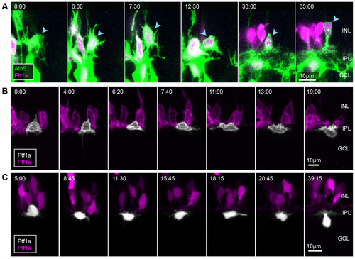
Basally migrating RINs in apical regions of the retina appear bipolar. (A) Selected frames from a movie of a Ptf1a:DsRed retina starting at ~45hpf. Images are shown as maximum intensity z-projections. At 0min, tips of RIN apical processes (arrowheads) are seen to be attached to the apical surface of the retina (top dashed green line). At 540min, when dACs (blue arrows) can be seen to separate from the main body of RINs in the middle of the retina, multipolar RINs, presumably HCs, can be seen migrating towards the OPL (orange arrows). (B) Selected time frames from a movie of Ptf1a:DsRed cells transplanted into a WT embryo starting at ~45hpf. Blue arrowheads indicate a RIN that transitions from bipolar to multipolar morphology. AS, apical surface; BS, basal surface. |

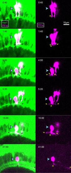
RINs transition directly into bipolar morphologies after birth. (A-C) Selected frames of movies starting at ~44hpf showing an HC, an iAC and a dAC (indicated by blue arrowheads) from birth. (D) Contour maps of the dAC shown in C at several time points. The dAC was traced z-slice by z-slice. Each trace was coloured based on the position of the z-slice relative to the centre of the cell. z-slices are spaced 1µm apart. Time is shown as hours relative to cell birth. Scale bars: 10µm. AS, apical surface; BS, basal surface; ONL, outer nuclear layer. |

HCs transition to flattened morphologies and migrate tangentially at regions near the future OPL. (A,B) Two examples of HCs dividing after migrating basally to the middle of the retina. Time shown in h:min relative to the start of the movie, ~50hpf. Blue arrowheads indicate the HC being tracked. Orange arrowheads indicate its sister after terminal division. |
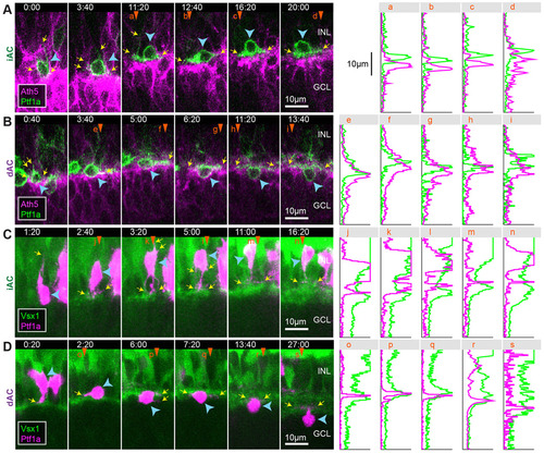
iACs and dACs undergo a stereotypical sequence of behaviours to polarize and migrate to their respective cell layers. (A) Example of an iAC (blue arrowhead) stratifying in the INL. (B,C) Two examples of dACs (white) migrating into the GCL. Times are shown in h:min relative to the start of the movies, ~48hpf. |

Early stages of IPL formation. (A) Snapshot of a 55hpf retina showing different stages of retinal development along the developmental wave. The RIN labelled with the orange asterisk probably represents a misplaced cell. (B) Quantification of the degree of interdigitation along the apical surface of RGCs, drawn as a white line in A. The vertical grey line indicates the angular position of the minimum value, and the corresponding 20° segment is indicated by the yellow line and yellow arrowheads in A. (C) The average line intensity values of the boxed region in A, showing the BC plexus (Crx) located between the RGC (Ath5) dendritic plexus and RINs (Ptf1a) in this region. (D) Selected frames of a movie of an RGC (blue arrowheads) migrating basally past RINs (orange arrowheads) and stratifying at a location basal to the RINs. (E) Selected frames of a movie of a BC axon (blue arrowheads) retracting to the basal side of RINs in the INL before the subsequent migration of a dAC (orange arrowheads) into the GCL. Time is shown as h:min from the start of the imaging sessions, at ~44hpf (D) and ~48hpf (E). |
iACs and dACs differentially stratify within the proto-IPL. (A,B) Selected frames of a movie of an iAC (A) and a dAC (B) transitioning from multipolar morphology to unipolar morphology in the Ath5 background. (C,D) Selected frames of a movie of an iAC (C) and a dAC (D) transitioning from multipolar morphology to unipolar morphology in the Vsx1 background. Panels on the right indicate the intensity line profile of the corresponding vertical line marked by orange arrowheads. Yellow arrows indicate the tips of the cells′ processes. Blue arrowheads indicate cell somas. Time is shown as h:min from the start of the imaging sessions, at ~50hpf. |
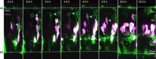
Occasionally, RINs are born near the region of the proto-OPL Selected frames from a movie of an atypical RIN born near the region of the proto-OPL. Movie starts at ~48 hpf. |

iACs and dACs undergo tangential migration near the proto-IPL A. Example of iAC (white) migrating tangentially about half a somal length via a process extending laterally from the basal side of the cell. B. Example of a dAC (white) migrating tangentially via process extending from its flattened soma as it migrates basally into the GCL. Times are shown in h:min relative to the start of the movies. Movies start at ~48hpf. |

Example of an atypical dAC migrating into the GCL Selected frames from a movie of a dAC (orange arrowheads) migrating into the GCL later than usual, when RGCs in the same clone are already clearly stratified. The dAC does not flatten, but squeezes through the forming IPL rapidly. For comparison, a dAC that migrated earlier is also indicated (blue arrowheads). Movie starts at ~48 hpf. |
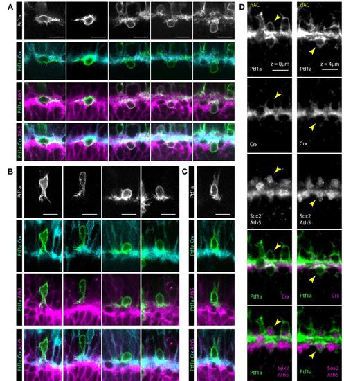
iACs and dACs differentially stratify in the proto-IPL A. Isolated labelled dACs identified in 48-60 hpf SoFa2 retinas, where RINs are mosaically labelled with Ptf1a driven YFP, BCs are labelled using Crx driven CFP and RGCs are labelled using Ath5 driven RFP. Each column shows a different presumptive dAC. Images are arranged from left to right to suggest a possible sequence of migration. YFP labelled cells found basal to the BC plexus generally run lateral processes across the interface of the BC (cyan) plexus and the RGC plexus (magenta). B. Most isolated labelled iACs identified in the SoFa2 retina appear to initially stratify in the apical side of the BC plexus. Images are arranged from left to right to suggest a possible sequence of migration. C. Rarely, iACs appear to stratify in the middle of the BC plexus. Images are arranged from left to right to suggest a possible sequence of migration. D. The SoFa2 retina for starburst ACs using Sox2 antibody. A starburst iAC and a starburst dAC close to each other are seen to stratify on the apical side of the BC plexus, and at the interface between the BC plexus and RGC plexus, respectively. |
Example of a iAC stratifying in the middle of the BC plexus |