
Image
|
Figure Caption
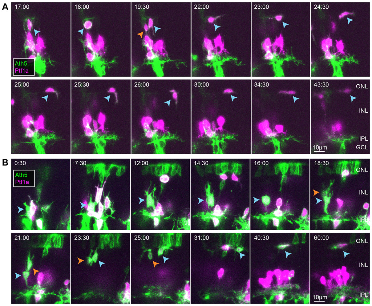
Fig. 3
HCs transition to flattened morphologies and migrate tangentially at regions near the future OPL. (A,B) Two examples of HCs dividing after migrating basally to the middle of the retina. Time shown in h:min relative to the start of the movie, ~50hpf. Blue arrowheads indicate the HC being tracked. Orange arrowheads indicate its sister after terminal division.
Acknowledgments
This image is the copyrighted work of the attributed author or publisher, and
ZFIN has permission only to display this image to its users.
Additional permissions should be obtained from the applicable author or publisher of the image.
Full text @ Development