
FIGURE
Fig. S6
Fig. S6
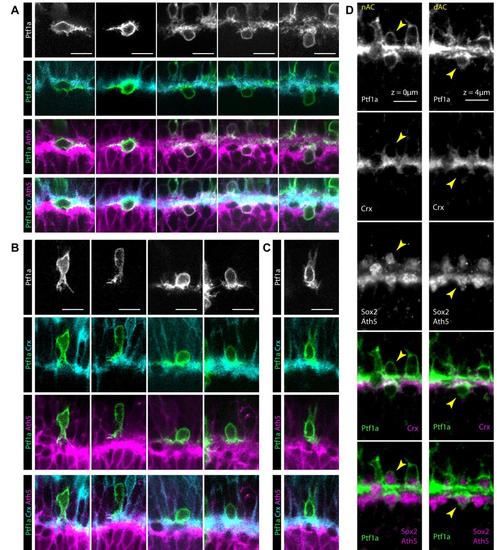
iACs and dACs differentially stratify in the proto-IPL A. Isolated labelled dACs identified in 48-60 hpf SoFa2 retinas, where RINs are mosaically labelled with Ptf1a driven YFP, BCs are labelled using Crx driven CFP and RGCs are labelled using Ath5 driven RFP. Each column shows a different presumptive dAC. Images are arranged from left to right to suggest a possible sequence of migration. YFP labelled cells found basal to the BC plexus generally run lateral processes across the interface of the BC (cyan) plexus and the RGC plexus (magenta). B. Most isolated labelled iACs identified in the SoFa2 retina appear to initially stratify in the apical side of the BC plexus. Images are arranged from left to right to suggest a possible sequence of migration. C. Rarely, iACs appear to stratify in the middle of the BC plexus. Images are arranged from left to right to suggest a possible sequence of migration. D. The SoFa2 retina for starburst ACs using Sox2 antibody. A starburst iAC and a starburst dAC close to each other are seen to stratify on the apical side of the BC plexus, and at the interface between the BC plexus and RGC plexus, respectively. |
Expression Data
Expression Detail
Antibody Labeling
Phenotype Data
Phenotype Detail
Acknowledgments
This image is the copyrighted work of the attributed author or publisher, and
ZFIN has permission only to display this image to its users.
Additional permissions should be obtained from the applicable author or publisher of the image.
Full text @ Development