
- Title
-
Transgenic Zebrafish Recapitulating tbx16 Gene Early Developmental Expression
- Authors
- Wells, S., Nornes, S., and Lardelli, M.
- Source
- Full text @ PLoS One

GFP expression in stable transgenic lines consistent with tbx16 expression in wild type embryos. (a) At 12 hpf strong expression is seen in the polster (arrowhead), and the presomitic mesoderm (p). Expression persists in mesoderm that has formed somites (s). (b) At 24 hpf GFP expression is seen in the hatching gland (arrowhead) and in posterior somites, while it has faded from the earliest formed somites. (c) At 48 hpf expression remains in the hatching gland that has formed from the polster (arrowhead) and persists in the posterior somites. (d) The same embryo as in (c). GFP expression is observed throughout the notochord (n) highlighting cell extremities. Lateral views of embryos, anterior is left and dorsal is up. Scale bars in a–c = 250 μm, in d = 100 μm. EXPRESSION / LABELING:
|

Expression unique to individual stable transgenic lines. (a) Line 192A at 48 hpf. GFP expression is observed in regions of the midbrain (arrowhead) and in the epiphysis (e). Line 512B at 24 hpf (b) and 48 hpf (c). Expression is seen in the dorsal aorta and in ventral neurons (n) of the spinal cord. Line 812A at 24 hpf (d–e) and at 48 hpf (f). Expression can be seen in the epiphysis, the midbrain and in a banding pattern in rhombomeres (boxed). White box indicates the area enlarged in (e). Later expression is observed in the floor plate of the spinal cord (f). (g–h) Line 812C at 48 hpf. Expression is seen in the epiphysis and the midbrain. GFP is also noted in a specific subpopulation of spinal cord neurons and neurons posterior of the otic vesicle in the hindbrain (boxed). White box indicates the area enlarged in (h). Lateral views of embryos, anterior is left and dorsal is up. Scale bars in a, d, g = 250 μm, in b, c, e, f, h = 50 μm. |
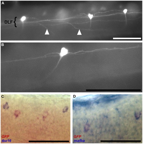
Identification of spinal cord neurons in line 812C at 48 hpf. (a) Lateral view of three GFP-positive neural cells situated in a non-uniform distribution along the dorsal longitudinal fasiculus (DLF) indicated by a tight grouping of neuronal projections. Axons projecting from neurons located in the contralateral DLF can be seen to rise from the ventral midline before joining the in-focus DLF (arrowheads). (b) Lateral view of a single GFP-positive neuron illustrating the projections originating from the cell. Two projections extend within the DLF – one ascending and one descending – and a third ventrally extending axon turns anteriorly and fades from view as it approaches the ventral midline before ascending in the contralateral DLF (not shown). (c) Two-colour in situ transcript hybridisation indicating that GFP-positive cells (stained red) are not the tbx16-positive dorsal longitudinal ascending interneurons (stained blue). (d) Two-colour in situ transcript hybridisation showing that neurons expressing GFP (stained red) also express mafba (stained blue). All embryos are positioned anterior left and dorsal up. Scale bars indicate 100 μm. |

Unillustrated author statements |