
- Title
-
Defective adult oligodendrocyte and Schwann cell development, pigment pattern, and craniofacial morphology in puma mutant zebrafish having an alpha tubulin mutation
- Authors
- Larson, T.A., Gordon, T.N., Lau, H.E., and Parichy, D.M.
- Source
- Full text @ Dev. Biol.

puma mutant adult zebrafish exhibit defects in pigment pattern, craniofacial morphology, Schwann cell development, and locomotor behavior. (A, A′) Adult stripes and craniofacial morphology are disrupted in puma mutants. (B, B′) Clearing and staining to reveal bone (red) and cartilage (blue) shows thinner dysmorphic bones of the skull in puma mutants (arrowheads) and ventral displacement of the jaw (arrow). (C, C′) External anatomy of head for wild-type and a severely affected puma mutant. (D–F) Development of Schwann cell defects. (D, D′) Early larvae exhibit mbp+ Schwann cells covering the lateral line nerve (arrow) in both wild-type and puma mutants (here, 4.0 mm standardized standard length, 4.0 SSL). (E, E′) During later post-embryonic development, mbp+ cells are more sparsely arranged in puma mutants (here, 6.3 SSL). (F, F′) mbp+ cells are nearly absent in puma mutants, with only rare residual cells found along the lateral line (arrow in F′); mbp+ cells found along dorsoventrally oriented nerves in wild-type (arrowheads in F) are completely lacking in puma (8.0 SSL). (G) Quantitative analysis of craniofacial defects in puma mutants. Shown are the proportions of individuals placed into different categories for severity of craniofacial defect, divided by sex and genotype. Numbers of individuals examined are given above the bars. Contingency table analysis shows that homozygous puma mutants were more likely than wild-type to exhibit craniofacial defects and such defects were more prevalent among males than females (genotype: X2 = 93.0, P < 0.0001; sex: X2 = 43.7, P < 0.0001; n = 171). Scale bars: in A, 4 mm for A, A′. In B, 1 mm for B, B′ in D, 60 μm for D, D′ in E, 60 μm for E, E′ in F, 60 μm for F, F′. EXPRESSION / LABELING:
|
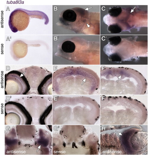
tuba8l3a is not spatially restricted in the embryo but is expressed most prominently in the central nervous system during post-embryonic development. Shown is staining with antisense and sense probes (diluted to equal concentrations) targeted to the 52 untranslated region of tuba8l3a, (A, A′) Widespread expression of tuba8l3a at 24 hpf. (B, B′) Lateral views of larvae (7.2 SSL) showing tuba8l3a transcript in the brain (arrow) and cranial ganglia (arrowhead). (C, C′) Dorsal views of the same individuals, illustrating tuba8l3a mRNA in the brain (arrow). Larvae in B–C are homozygous nacre mutants that lack otherwise obscuring melanophores due to an autonomously acting mutation in the mitfa transcription factor. Expression in wild-type larvae was indistinguishable from nacre mutants (not shown). (D, D′) tuba8l3a transcript is detectable in the inner nuclear layer of the retina (arrow). (E, E′, F, F′) More posteriorly, tuba8l3a is expressed in the periventricular grey zone of optic tectum. (G, G′, H) tuba8l3a staining (arrow) in the cranial ganglia. Larvae in D–H are 8–9 SSL. EXPRESSION / LABELING:
|
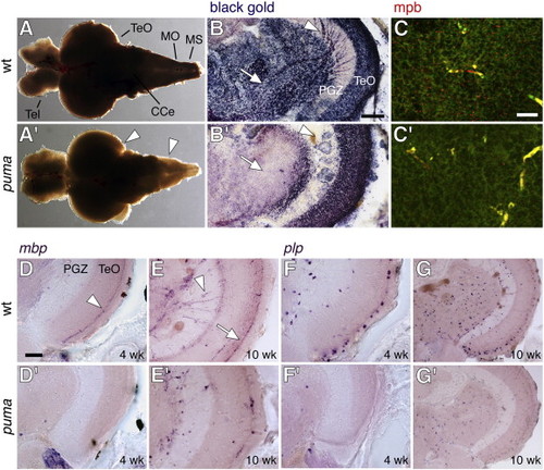
Deficient central nervous system myelination in puma mutant adults and juveniles. (A, A′) Decreased opacity of adult brain from puma as compared to wild-type; this difference is especially notable at the lateral edges and posteriorly (arrowheads). Anterior to the left. Tel, telencephalon; TeO, tegmentum opticum; CCe, corpus cerebelli; MO, medulla oblongata; MS, medulla spinalis. (B, B′) Black Gold II staining for myelin in transverse sections of wild-type and puma mutant adults. Myelin is stained blue-black whereas other tissue is purple-red. Reduced staining in puma is evident to some degree within the optic tectum (TeO) but is more pronounced in ventral–medial regions (arrow), internal to the periventricular grey zone (PGZ). Radially oriented black gold staining (arrowhead) also is largely absent from the PGZ itself of the puma mutant. (C, C′) Antiserum against zebrafish mbp (red) stains numerous punctate foci in the ventral–medial brain of wild-type but far fewer in the corresponding region of puma mutants. Green, background tissue fluorescence. Yellow, fluorescence of blood vessels in green and red channels. (D, D′–G′, G) In situ hybridization for mbp and plp in juveniles and adults (13 SSL, 24 SSL). (D, D′) In juveniles, staining for mbp is pronounced in a band (arrowhead) near the outer surface of the optic tectum (TeO) in wild-type (D) but is not apparent in puma (D′). (E, E′) In adults, abundant mbp transcripts are found radially in the PGZ (arrowhead) and at the outer surface of the optic tectum (arrow) in wild-type but are found at lower levels in puma. (F, F′) In juveniles, plp staining is detected in individual cell bodies both interiorly (arrowhead) and near the tectum surface (arrow) of wild-type but there are far fewer of these cells in puma mutants. (G, G′) In adults, numerous plp-expressing cell bodies are observed in wild-type, whereas in puma mutants there are fewer cells and those present are less densely stained. Scale bars: in B, 100 μm; in C, 20 μm; in D, 100 μm for D–G. EXPRESSION / LABELING:
PHENOTYPE:
|
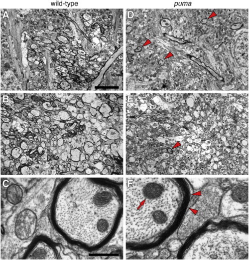
Reduced myelination in puma mutant brains revealed by transmission electron microscopy. (A–C) Representative regions of wild-type at low magnification (5000×, A and B) and higher magnification (60,000×, C) show numerous well-myelinated axon tracts. (D–F) Corresponding regions and magnifications in puma reveal far fewer myelinated axon tracts (arrowheads in D and E) though layering of individual myelin sheaths that are present is indistinguishable from wild-type (arrowheads in F; arrow, mitochondrion). Scale bars: in A, 5 μm, for A, B, D, E; in C, 500 nm for C, F. PHENOTYPE:
|

Defects in oligodendrocyte number and patterning during the larval-to-adult transformation of puma mutants. (A, A′, B, B′) Wild-type zebrafish larvae exhibit many more plp+ oligodendrocytes in the brain than do puma mutants both during the middle larval period (A, A&prime& ~ 6.5 SSL) and the late larval period (B, B&prime& ~ 8.0 SSL). (C, C′, D, D′, E, E′) mbp expression also differs between wild-type and puma mutants during middle and later larval development (C, C′ and D, D′, respectively); a higher magnification view of different larvae is shown in E, E′. Note especially the absence of most myelinated fibers in the puma mutant and the concentration of mbp mRNA in cell bodies. (F, F′, G, G′) The early oligodendrocyte marker olig1 does not differ in expression between genotypes at middle or later larval stages, nor do several additional markers of particular cell lineages or activities (H–J; see text for details). Scale bars: in A, 100 μm, for A, A′, B, B′, C, C′, D, D′, F, F′. In E, 40 μm for E, E&prime& in G, 100 μm for G, G′ in H, 100 μm for H, H&prime& in J, 100 μm for I, J&prime& in J, 100 μm for J, K′. EXPRESSION / LABELING:
PHENOTYPE:
|

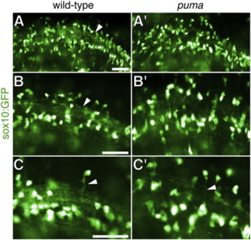
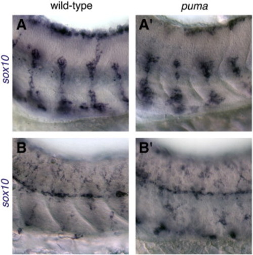
Defects in oligodendrocyte patterning and myelination in puma mutant early larvae. (A–C) sox10:GFP transgene reveals oligodendrocyte cell bodies and processes, that are more disorganized in puma mutants than in wild-type at 5 dpf. Cell bodies are deliberately overexposed (shown here in white) to reveal fainter processes. (A, A′) Low magnification showing the orderly array of myelinating processes in wild-type (arrow), and more disorganized processes in puma mutants. (B, B′) Higher magnification of different individuals. (C, C′) Relatively long processes are observed in both wild-type and puma mutants (arrowheads). Scale bars: in A, 40 μm for A, A′; in 40 μm for B, B′; in C, 40 μm for C, C′. |

Unillustrated author statements EXPRESSION / LABELING:
PHENOTYPE:
|
Reprinted from Developmental Biology, 346(2), Larson, T.A., Gordon, T.N., Lau, H.E., and Parichy, D.M., Defective adult oligodendrocyte and Schwann cell development, pigment pattern, and craniofacial morphology in puma mutant zebrafish having an alpha tubulin mutation, 296-309, Copyright (2010) with permission from Elsevier. Full text @ Dev. Biol.