
- Title
-
SoxB1 transcription factors restrict organizer gene expression by repressing multiple events downstream of Wnt signalling
- Authors
- Shih, Y.H., Kuo, C.L., Hirst, C.S., Dee, C.T., Liu, Y.R., Laghari, Z.A., and Scotting, P.J.
- Source
- Full text @ Development

Sox3 inhibits gastrulation and represses markers of the zebrafish organizer and mesoderm. (Aa-c′) Injection of sox3 mRNA at the 1- to 2-cell stage (Aa′) caused disruption of the blastoderm/yolk cell border at 6.5 hpf (asterisk). At 9 hpf, sox3-injected embryos (Ab′) showed delayed gastrulation and a thickening on one side (asterisk) with a pronounced ventralised phenotype in those that survived to 24 hpf (Ac′). (Ba-e′) Injected sox3 caused a dramatic reduction in the expression of sqt, gsc, chd and nog1 and a substantial gap in the ring of ntl expression, which was coincident with the region of injected sox3, as shown by staining for HA (brown) (Bd′, inset). (C) Compared with wild-type (WT) (a-e), injection of the dominant-negative forms of Sox3, Sox3N40I (f-j) and Sox3dNLS (k-o), did not repress the endogenous domains of expression of sqt, gsc, chd, ntl or nog1, but caused ectopic expression in the majority of embryos (arrowheads). The numbers of embryos showing ectopic expression/number injected are shown bottom right. The period of development is shown below each panel. Gene expression was analysed by whole-mount in situ hybridisation (blue). Animal pole views, except for A which was viewed laterally. Dorsal is to the right, except for Aa, Aa′, Bd, Bd′, for which orientation cannot be certain. Scale bars: ∼200 μm. |

Dominant-negative Sox3 constructs induce ectopic expression of bozozok and axis duplication. (A) Effect of Sox3 mutants as compared with WT Sox3 (a) on boz expression by RNA injection at the 1- to 2-cell stage and analysed at 4.5 hpf. Animal pole view, dorsal to the right, except Ab for which orientation cannot be certain. Injection of WT Sox3 caused loss of boz expression (b). Neither Sox3N40I (c) nor Sox3dNLS (d) repressed endogenous boz expression, but caused ectopic expression of boz in the majority of embryos. (B) Analysis 24 hours after injection showed that Sox3dNLS caused various extents of axis duplication, as shown by analyzing the expression of ncad (cdh2) (a-a′), hoxb1b (b-b′), shh (c-c′) and krox20 (egr2) (d-d′). (C) The percentage of embryos of each duplication phenotype following injection of Sox3dNLS RNA (n=279). Scale bars: ∼200 μm. |
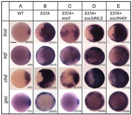
Sox3 inhibits Wnt/β-catenin signalling. WT Sox3 inhibited the ability of S37A β-catenin 2 to activate the expression of boz, sqt, gsc and chd in vivo (A-C). Neither the dNLS (D) nor N40I (E) mutants of Sox3 were able to inhibit this response to S37A RNA. Animal pole views, dorsal right. Scale bar: ∼200 μm. |

The effects of Sox3dNLS require Wnt/β-catenin signalling. (A-G) Ectopic activation of boz, sqt, gsc and chd by Sox3dNLS RNA injected at the 1- to 2-cell stage and assayed at 4.5 hpf (B, compare with uninjected WT embryo, A) was blocked by WT Sox3 (C), but also by dnTcf3 (D,E) or a β-catenin 2 MO (F,G). Animal pole views, dorsal right. Scale bar: ∼200 μm. |
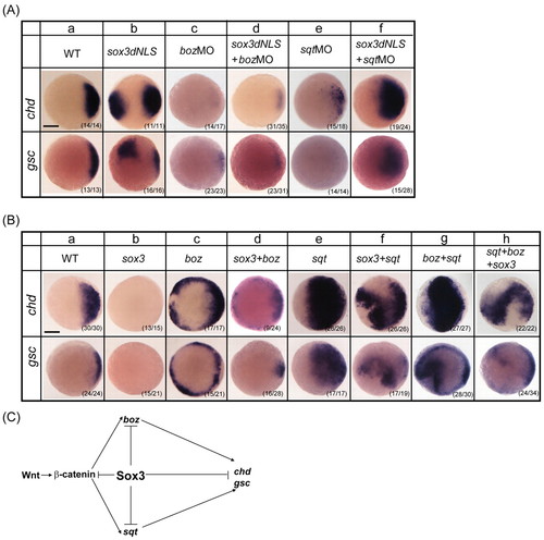
Interaction between Sox3, Boz and Sqt. Animal pole views, dorsal to the right. (A) Both endogenous (a) and ectopic expression of gsc and chd induced by Sox3dNLS RNA injected at the 1- to 2-cell stage (b) was blocked by both boz (c,d) and sqt (e,f) MOs. (B) Compared with uninjected WT embryos (a), injection of WT sox3 RNA at the 1- to 2-cell stage and assayed at 4.5 hpf repressed expression of gsc and chd (b), whereas boz and/or sqt activated their expression (c,e,g). Co-injection of sox3 RNA with boz (d) or sqt (f) or both (h) RNAs resulted in intermediate levels of gsc and chd expression. (C) Model of Sox3 function in pathways that promote organizer formation. T-bars indicate inhibition and arrows indicate requirement for activation. Scale bars: ∼200 μm. |
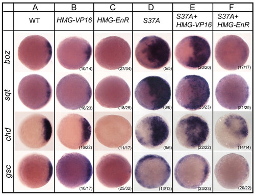
HMG-EnR constructs mimic the effects of WT Sox3 on organizer markers. All embryos are 4.5 hpf, animal pole view, dorsal right. Sox3HMG-EnR (C), but not Sox3HMG-VP16 (B), repressed all markers (boz, sqt, chd, gsc), as compared with controls (A). Similarly, Sox3HMG-EnR (F), but not Sox3HMG-VP16 (E), inhibited the activation of these markers by S37A β-catenin 2 (D). Scale bar: ∼200 μm. |
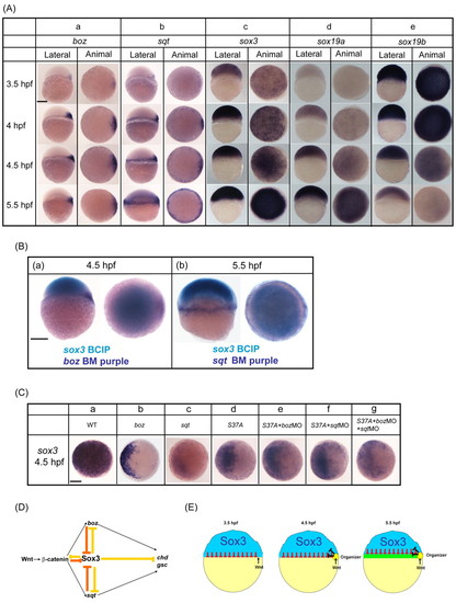
Reciprocal expression and transcriptional repression between Sox3, β-catenin 2, Boz and Sqt. (A) Lateral and animal views, dorsal right, showing endogenous expression of boz (a), sqt (b), sox3 (c), sox19a (d) and sox19b (e). (B) Double in situ hybridisation for boz/sox3 (a) or sqt/sox3 (b). sox3 is detected with BCIP (pale blue), boz/sqt with BM purple. (C) Effect of S37A, Boz and Sqt on sox3 expression. Animal view of sox3 expression at 4.5 hpf, dorsoventral orientation cannot be certain. Endogenous sox3 expression (a) was repressed by injection of boz (b), sqt (c) or S37A β-catenin 2 (d) RNA. MOs directed against boz and/or sqt did not affect the ability of S37A β-catenin 2 RNA to inhibit the expression of sox3 (e-g). (D) Model for the role of Sox3 in restricting organizer formation. T-bars indicate mutual repression between Sox3 and β-catenin 2, boz and sqt. (E) Taking our experimental data together with the endogenous domains of expression illustrated above, this leads us to a model in which at 4-4.5 hpf, high levels of Wnt signalling on the dorsal side (yellow) together with vegetal Nodal signalling inhibit sox3 expression and induce expression of boz, which can also repress the expression of sox3 in the dorsal margin. By 5.5 hpf, sox3 is further depleted throughout the whole margin by Nodal signalling, allowing mesendoderm to form (green). Away from the margin, in the prospective ectoderm, Nodal signalling is too weak to repress sox3 expression and so Sox3 remains present (blue), inhibiting the expansion of mesoderm and organizer into this region (red bars). Scale bars: ∼200 μm. |

Comparison of the effects of Sox3, Sox19a and Sox19b on the expression of organizer genes. All embryos are at 4.5 hpf; animal pole view, dorsal to the right. Compared with uninjected WT embryos (A), overexpression of Sox3 (B), Sox19a (C) or Sox19b (D) resulted in the loss or reduction of expression of boz, sqt, chd and gsc. Like Sox3 (F), Sox19b (H) was largely able to rescue the effect of Sox3dNLS (E), whereas Sox19a was not (G). |
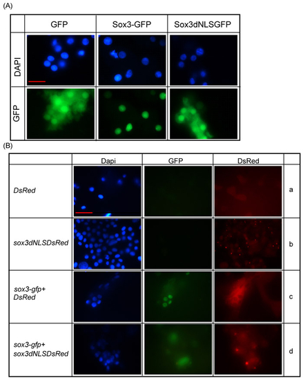
Sox3dNLS disrupts nuclear localisation. (A) GFP-tagged WT Sox3 expressed from RNA injected at the 1- to 2-cell stage was almost exclusively located in the nucleus of zebrafish embryo cells, whereas the distribution of a Sox3dNLS-GFP fusion appeared identical to GFP alone, as seen in both the nucleus and cytoplasm. (Ba-d) Sox3dNLS-dsRED and dsRED alone showed similar distribution throughout the cell. When co-expressed, Sox3dNLS-dsRED caused a loss of nuclear localisation of WT Sox3-GFP, whereas dsRED alone did not (compare c with d). Scale bar: ∼10 μm. |