
Image
|
Figure Caption
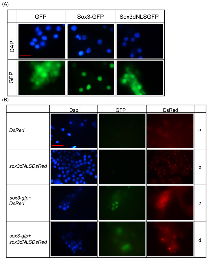
Fig. S1 Sox3dNLS disrupts nuclear localisation. (A) GFP-tagged WT Sox3 expressed from RNA injected at the 1- to 2-cell stage was almost exclusively located in the nucleus of zebrafish embryo cells, whereas the distribution of a Sox3dNLS-GFP fusion appeared identical to GFP alone, as seen in both the nucleus and cytoplasm. (Ba-d) Sox3dNLS-dsRED and dsRED alone showed similar distribution throughout the cell. When co-expressed, Sox3dNLS-dsRED caused a loss of nuclear localisation of WT Sox3-GFP, whereas dsRED alone did not (compare c with d). Scale bar: ∼10 μm.
Acknowledgments
This image is the copyrighted work of the attributed author or publisher, and
ZFIN has permission only to display this image to its users.
Additional permissions should be obtained from the applicable author or publisher of the image.
Full text @ Development