
- Title
-
Extracellular divalent cations modulate aminoglycoside-induced hair cell death in the zebrafish lateral line
- Authors
- Coffin, A.B., Owens, K.N., Raible, D.W., and Rubel, E.W.
- Source
- Full text @ Hear. Res.

Neuromast hair cells have a minimal calcium requirement. Fish were incubated for 90 min in embryo medium with no added calcium or in embryo medium with moderate (900 μM) calcium (standard embryo medium). (A) SO2 neuromast from a 5 dpf fish incubated in medium with no added calcium (nominal Ca2+-free; left) or in normal embryo medium (right). Hair cell nuclei are labeled with YO-PRO-1 (blue) and hair cell cytoplasm with FM 1-43 (red). Arrows in the left panel point to condensed nuclei. Scale bar = 5 μm and applies to both panels. (B) Hair cells were counted in four neuromasts per fish and hair cell numbers were separately normalized to the number of hair cells in control neuromasts. The normalized average for all four neuromasts is shown here. N = 9–10 fish/treatment, bars are mean ± 1 SD. Hair cell number is significantly different between the two conditions (unpaired t-test, p ≤ 0.01). |
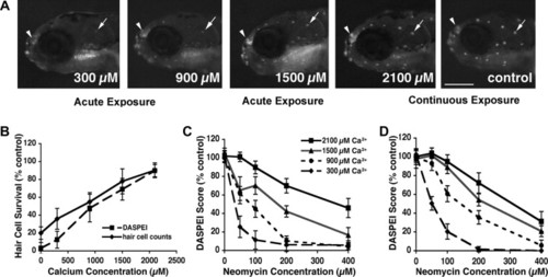
Calcium protects hair cells from neomycin in a dose-dependent manner. (A) Fish were acutely exposed to 100 μM neomycin in embryo medium with variable Ca2+ concentration and hair cell survival was assessed with the vital dye DASPEI. The arrow indicates the MI1 neuromast in each image as a reference, while the arrowhead points to the olfactory epithelium, which serves as an internal labeling control. Numbers on each image indicate the Ca2+ concentration. The scale bar in the “control” image is 0.5 mm. (B) Mean hair cell survival following acute 100 μM neomycin exposure was assessed using DASPEI scoring (dashed line) from 10 neuromasts per fish or by direct counts of YO-PRO-1-labeled hair cells (solid line) from four neuromasts per fish. Mock-treated controls are not shown but are represented by the 100% level. Increased calcium shows significant protection against neomycin at this dose (two-way ANOVA, p < 0.001) (C and D). Dose-response curves for acute (C) and continuous (D) neomycin exposure in embryo medium containing either 300, 900, 1500, or 2100 μM Ca2+. Mean hair cell survival significantly increases with increasing external Ca2+ for either exposure time course (p < 0.001). The legend in panel C applies to both C and D. N = 9–16 fish/treatment, error bars are ±1 SD. |
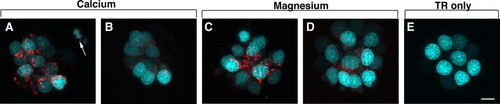
GTTR uptake is substantially antagonized by external calcium and to a lesser degree by magnesium. Fish were incubated for 10 min in GTTR diluted in (A) 300 μM calcium, (B) 2100 μM calcium, (C) 300 μM magnesium, or (D) 2100 μM magnesium. (E) Negative control neuromast from a fish incubated with Texas Red only. GTTR fluorescence is red; nuclei are counter-stained blue with YO-PRO-1. GTTR fluorescence is greatly reduced in the presence of high calcium but there is little difference in fluorescence between the low and high magnesium conditions. The arrow in A indicates a fragmented hair cell. The scale bar in E is 5 μm and applies to all panels. |
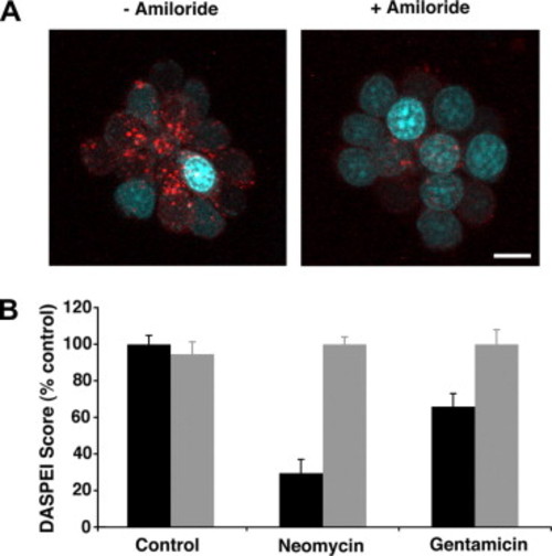
Amiloride blocks drug uptake and the resulting loss of hair cells. (A) Amiloride attenuates short-term (10 min) GTTR uptake (right) as compared to GTTR uptake without amiloride (left). Hair cell nuclei are labeled with YO-PRO-1 (blue), scale bar = 5 μm and applies to both panels. (B) 1 mM amiloride prevents hair cell loss from acute neomycin or gentamicin exposure (unpaired t-tests, p > 0.05 for amiloride + aminoglycoside as compared to mock-treated controls). Black bars = no amiloride, gray bars include 1 mM amiloride. Controls show that amiloride alone does not alter DASPEI scores (unpaired t-test, p > 0.05). Error bars in (B) are ±1 SD. All experiments shown here were conducted in standard EM (∼1 mM Ca2+). |
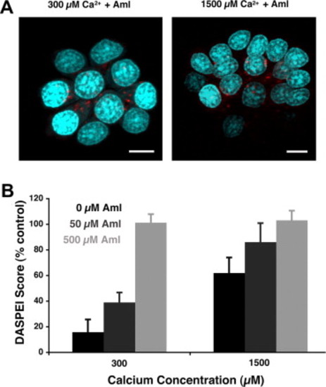
Interplay of amiloride and calcium in aminoglycoside uptake and hair cell death. (A) 50 μM amiloride reduces GTTR uptake under low calcium (300 μM) conditions (left) but does not completely prevent GTTR uptake even under higher (1500 μM) calcium conditions (right). Hair cell nuclei are labeled with YO-PRO-1 (blue), scale bars are 5 μm. (B) Amiloride increases hair cell survival proportionally regardless of calcium concentration. Black bars represent the effect of calcium only on hair cell loss for two concentrations of calcium (300 or 1500 μM). Dark gray bars depict hair cell survival in the indicated calcium concentration with 50 μM amiloride added, while light gray bars represent hair cell survival with 500 μM amiloride. Error bars are ±1 SD. |
Reprinted from Hearing Research, 253(1-2), Coffin, A.B., Owens, K.N., Raible, D.W., and Rubel, E.W., Extracellular divalent cations modulate aminoglycoside-induced hair cell death in the zebrafish lateral line, 42-51, Copyright (2009) with permission from Elsevier. Full text @ Hear. Res.