
- Title
-
Two deltaC splice-variants have distinct signaling abilities during somitogenesis and midline patterning
- Authors
- Mara, A., Schroeder, J., and Holley, S.A.
- Source
- Full text @ Dev. Biol.
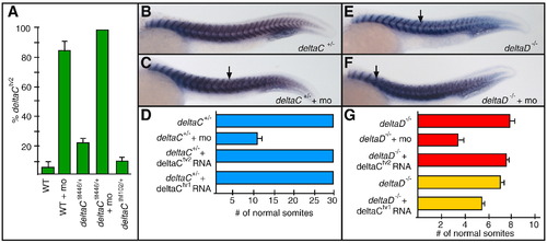
deltaCtv2 does not signal effectively during somitogenesis. (A) qPCR showing the relative abundance of deltaCtv2 in 15 somite embryos of various genetic backgrounds. Levels of deltaCtv2 increase significantly in morpholino (mo) injected embryos. The amount of deltaCtv2 mRNA is graphed as a percentage of total deltaC mRNA. (B–C) titin staining marks the somite boundaries of deltaC+/- embryos. The first disrupted somite border in a morpholino injected embryo is indicated by the arrow in (C). (D) The average number of somites formed in each experimental condition. n = 93, 102, 73, 115 from top to bottom. (E–F) The onset of border defects in deltaD-/- embryos is shifted anteriorly in morpholino injected embryos. Arrows indicate the first abnormal border. (G) The average number of somites formed in each experimental condition. n = 73, 91, 84, 139, 99 from top to bottom. Two different sets of mutant and sibling embryos were used for the experiments marked in red and yellow. Error bars indicate standard deviation. EXPRESSION / LABELING:
PHENOTYPE:
|
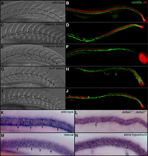
Over-expression of different deltas promotes hypochord cell fates over notochord at distinct positions along the anterior–posterior axis. (A–J) DIC and fluorescent in situ images of embryos with notochord to hypochord conversions at different positions along the anterior–posterior axis. DIC images are at 24 somites, while in situ images are at 24 hpf. ntl expression (red) marks the notochord, while αcol2a (green) shows the floor plate and hypochord. Embryos lacking notochord entirely are present, but have a twisted body axis and cannot be imaged easily via fluorescence, hence a weaker phenotype with some residual ntl expression is shown in (H). (K–M) Embryos at the 24 somite stage stained for αcol2a. Injection of some delta mRNAs can rescue hypochord formation in embryos where both deltaC and deltaD function have been impaired (i.e. deltaC-/- embryos injected with a deltaD morpholino). Select hypochord cells are marked by arrows or brackets. |
Reprinted from Developmental Biology, 318(1), Mara, A., Schroeder, J., and Holley, S.A., Two deltaC splice-variants have distinct signaling abilities during somitogenesis and midline patterning, 126-132, Copyright (2008) with permission from Elsevier. Full text @ Dev. Biol.