
- Title
-
Expression and Regulation of the Vitamin D Receptor in the Zebrafish, Danio rerio
- Authors
- Craig, T.A., Sommer, S., Sussman, C.R., Grande, J.P., and Kumar, R.
- Source
- Full text @ J. Bone Miner. Res.

Section of adult male zebrafish. Immunostaining with anti-VDR antibody or pre-immune serum. 50 X original magnification. Panel A. Sagittal section. Immunostaining with anti-VDR antibody of the intestine (I), Liver (L), pancreas (P), kidney (K and arrow), and testis (T). Panel B. Sagittal section. Pre-immune serum. Note absence of staining in the intestine, liver, pancreas, kidney and testis. Panel C. Sagittal section. Immunostaining with anti-VDR antibody of the gills (G), kidney (K), liver (L) and brain (B). Panel D. Transverse section. Immunostaining with anti-VDR antibody of gills (G), epithelial lining of oropharynx (O), and spinal cord (SC). Panel E. Sagittal section. Immunostaining with anti-VDR antibody of olfactory organ (OO) and eye (E). Panel F. Sagittal section. Pre-immune serum. Note absence of staining of olfactory organ (OO) and eye (E). The dark brown color represents the VDR. EXPRESSION / LABELING:
|

Section of adult male zebrafish. Immunostaining with anti-VDR antibody pre-adsorbed with recombinant Danio rerio full-length VDR. 50 X original magnification. Panel A. Intestine. Panel B. Liver and kidney. Panel C. Gills. Panel D. Testis. Panel E. Eye. Note absence of immunostaining in all tissues. |

Section of adult male zebrafish. Immunostaining with anti-VDR antibody or pre-immune serum. 200 X original magnification. Panel A. Sagittal section. Immunostaining with anti-VDR antibody of kidney. Note staining of glomerular epithelium (G) and tubular epithelium (T). Note absence of staining of vessels (V). Panel B. Sagittal section. Pre-immune serum. Note absence of staining of tubules glomeruli and blood vessels of the kidney. Panel C. Sagittal section. Immunostaining with anti-VDR antibody of gills. Note staining of epithelial cells in the gill lamellae (L). Also notes staining of chondrocytes in gill filaments (CC in F). Panel D. Sagittal section. Pre-immune serum. Note absence of staining of gill structures. Panel E. Sagittal section. Immunostaining with anti-VDR antibody. Note immunostaining of olfactory organ epithelium (OOE). Panel F. Sagittal section. Pre-immune serum. Note absence of staining of olfactory organ epithelium. Panel G. Sagittal section. Immunostaining with anti-VDR antibody. Note immunostaining of epithelial cells of the intestine (EC). Unstained cells are goblet cells. Panel H. Sagittal section. Pre-immune serum. Note absence of staining of epithelial cells of the intestine. EXPRESSION / LABELING:
|

Section of adult male zebrafish. Immunostaining with anti-VDR antibody or pre-immune serum. 200 X original magnification. Panel A. Sagittal section. Immunostaining with anti-VDR antibody of pancreas. Note staining of acinar cells of the pancreas (AC) and absence of staining of islets (I). Panel B. Sagittal section. Immunostaining with anti-VDR antibody of bone. Note staining of osteoblasts lining decalcified bone (Ob). Panel C. Sagittal section. Immunostaining with anti-VDR antibody of liver. Note immunostaining of hepatocytes (Hc) and cholangiocytes (Cc). Panel D. Sagittal section. Pre-immune serum. Note absence of staining of hepatocytes and cholangiocytes. Panel E. Sagittal section. Immunostaining with anti-VDR antibody. Note faint immunostaining of cardiac myocytes. The immuno-peroxidase staining within the ventricular cavity represents staining of erythrocytes whose endogenous peroxidase activity has not been completely suppressed. Panel F. Sagittal section. Pre-immune serum. Note absence of staining of cardiac myocytes. Panel G. Sagittal section. Immunostaining with anti-VDR antibody. Note immunostaining of Sertoli cells (Sc) of the testis. Panel H. Sagittal section. Pre-immune serum. Note absence of staining of Sertoli cells of the testis. EXPRESSION / LABELING:
|

Immunostaining of tissues from adult female zebrafish with anti-VDR antibody or pre-immune serum. 50 X original magnification. Panel A. Developing oocytes immunostained with anti-VDR antibody. Panel B. Immunostained with pre-immune serum. Panel C. The gills immunostained with anti-VDR antibody. Panel D. The gills immunostained with pre-immune serum. EXPRESSION / LABELING:
|

Immunostaining of the eye and neural tissues with anti-VDR antibody or pre-immune serum. 200 X original magnification. Panel A. Section of the retina immunostained with anti-VDR antibody. a = ganglion cell layer; b = inner plexiform layer; c = inner nuclear layer; d = outer plexiform layer; e = outer nuclear layer; f = rods; g = pigmented layer. Panel B. Section of retina using pre-immune serum. Note absence of staining in all layers. Panel C. Section of brain immunostained with anti-VDR antibody. Panel D. Section of brain immunostained with pre-immune serum. Panel E. Section of brain immunostained with anti-VDR antibody (400 X). Panel F. Section of spinal cord staining with anti-VDR antibody. EXPRESSION / LABELING:
|
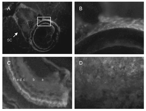
Cryosections of adult zebrafish labeled with VDR antibody visualized with Cy5 (red). Nuclei were labeled with DAPI (blue). A. Vertebra showing labeling in spinal cord (sc), vertebral body (boxed area), and surrounding muscle. B. Higher magnification of the vertebral body (boxed region). C, retina showing ganglion cell layer (a), inner plexiform layer (b), inner nuclear layer (c), outer plexiform layer (d), outer nuclear layer (e), and photo receptor layer (f). D, gills. EXPRESSION / LABELING:
|

Immunostaining of 48 hour post-fertilization zebrafish embryos with anti-VDR antibody or pre- immune serum. 100 X original magnification. Panel A. Coronal section. 48 hour post-fertilization embryo immunostained with anti-VDR antibody. Note staining (brown color) of cells within the eye (neural retina), the brain (diencephalon) and the developing mandible. 100 X original magnification. Panel B. Sagittal section. 48 hour post-fertilization embryo immunostained with immune serum. Note staining of cells of the neural retina, brain and developing mandible. Panel C. Coronal section. 48 hour post-fertilization embryo immunostained with pre-immune serum. Note absence of staining. 100 X original magnification. Panel D. Sagittal section. 48 hour post-fertilization embryo immunostained with pre-immune serum. Note absence of staining in cells of the neural retina, brain and developing mandible. EXPRESSION / LABELING:
|

Immunostaining of 96 hour post-fertilization zebrafish embryo with anti-VDR antibody or pre-immune serum. 100 X original magnification. Panel A. 96 hour post-fertilization embryo immunostained with anti-VDR antibody. Note staining (brown color) of cells within the eye (E), the brain (B) and the otic vesicle (O). 100 X original magnification. Panel B. 96 hour post-fertilization embryo immunostained with pre-immune serum. Note absence of staining for the VDR. 100 X original magnification. Panel C. Section of the eye immunostained with anti-VDR antibody; 200 X original magnification. L = lens; G = ganglion cells; R = photoreceptor cells; P = pigmented epithelial cells. Panel D. Otic vesicle immunostained with anti-VDR antibody. 200 X. original magnification. EXPRESSION / LABELING:
|
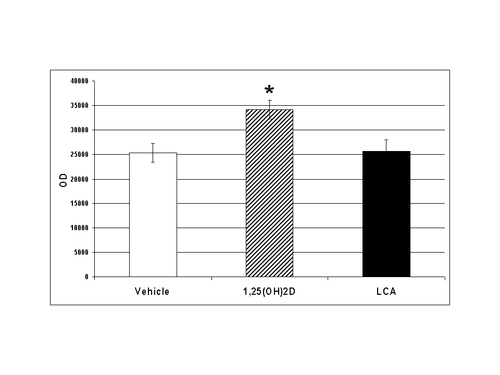
Relative concentrations of VDR in the intestine following treatment of adult male zebrafish with vehicle, 1α, 25(OH)2D3 or lithocholic acid (LCA). * p<0.05. Adult zebrafish were administered 25 ng of 1α, 25(OH)2D3 in 5 μl of propylene glycol, 25 ng of lithocholic acid in 5 μl of propylene glycol, or vehicle (5 μl of propylene glycol) parenterally. Twenty two hours later the fish were killed and tissues harvested as described. Equal amounts of protein (60 μg) were separated by SDS-PAGE using 10% acrylamide gels. Following electrophoresis, proteins were transferred to PVDF membranes and the VDR was detected by Western blotting methods as described. EXPRESSION / LABELING:
|