
- Title
-
The TORC2 Component, Sin1, Controls Migration of Anterior Mesendoderm during Zebrafish Gastrulation
- Authors
- Dumortier, J.G., David, N.B.
- Source
- Full text @ PLoS One
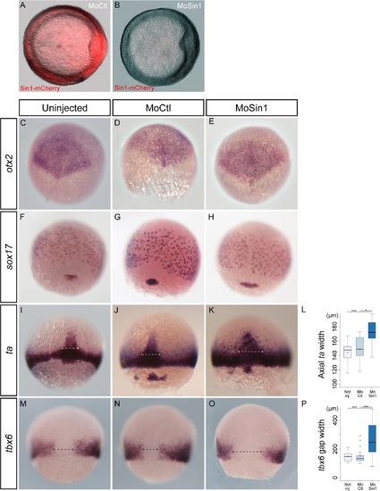
Loss of sin1 function leads to embryonic axis enlargement. A-B. At the dose used in this analysis, sin1 morpholino efficiently blocks translation of sin1 RNAs. A control morpholino (A) or a morpholino targeting the translation initiation site of sin1 (B) was co-injected at the 1-cell stage with mRNAs encoding a Sin1-mCherry fusion. Both images were acquired with the same exposure time. C-P. In situ hybridisations of genes expressed in the neuroectoderm (otx2), the endoderm (sox17), the mesoderm (ta) or the ventro-lateral mesoderm (tbx6). Embryos are either un-injected, injected with a control morpholino or with sin1 morpholino. They were fixed at mid-gastrulation. The three germ layers appear correctly formed. Mesoderm stainings nevertheless reveal a lateral widening of axial structures, quantified in L and P. EXPRESSION / LABELING:
PHENOTYPE:
|
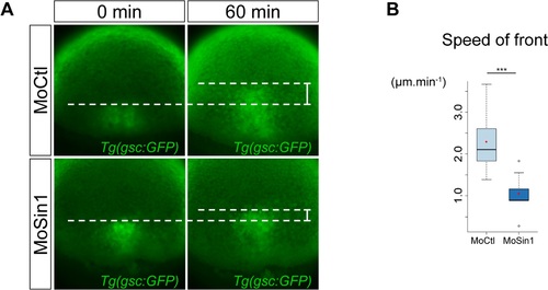
The prechordal plate migrates slower in sin1 loss of function. (A) Progression of the front of the prechordal plate was monitored in Tg(-1.8gsc:GFP) embryos (see S1 Movie). In embryos injected with the sin1 morpholino, the prechordal plate migrates twice slower than in embryos injected with a control morpholino. (B) Average speed of the front of the prechordal plate (distance travelled over one hour divided by 60 minutes), in embryos injected with a control morpholino or the sin1 morpholino. PHENOTYPE:
|

Absence of Sin1 affects protrusive activity of prechordal plate cells. (A) Diagram of the design of prechordal plate cell transplantation. Cells were labelled with Lifeact-mCherry and transplanted from shield to shield. (B) At 24 hpf, sin1 morphant cells transplanted into wild-type prechordal plates take part to the hatching gland. (C) Cells injected with Lifeact-mCherrry RNAs and either a control morpholino, or the sin1 morpholino, or CA-PI3K RNAs, or the sin1 morpholino and CA-PI3K RNAs, or the sin1 morpholino and CA-Rac1 RNAs were transplanted from shield to shield in Tg(-1.8gsc:GFP) embryos. Transplanted cells are within the prechordal plate (assessed by GFP expression), and contour of the plate is delineated (white dotted line). Orientations of their cytoplasmic extensions were measured relative to the animal pole and plotted as histograms (C) and cumulative plot in (D). (E) Frequencies of cytoplasmic extensions. |
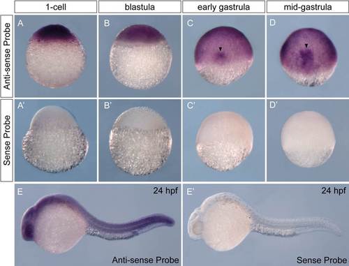
sin1 is expressed ubiquitously during the first 24 hours of development. In situ hybridisation of sin1 probe (A-E), or a sense probe used as a control (A′-E′), at the 1-cell stage (A), in blastula (sphere stage; B), at the onset of gastrulation (shield stage; C), at mid-gastrulation (75% epiboly; D) and at 24-hpf (E). sin1 appears maternally inherited and ubiquitously expressed. A stronger signal is observed in the forming embryonic axis during gastrulation (black arrowheads in C and D) |