
- Title
-
Expression of the fras1/frem gene family during zebrafish development and fin morphogenesis
- Authors
- Gautier, P., Naranjo-Golborne, C., Taylor, M.S., Jackson, I.J., and Smyth, I.
- Source
- Full text @ Dev. Dyn.

Expression of frem1a and frem2a during development. Expression of frem1a (A-I) and frem2a (J-Q). A,B: Lateral views of frem1a expression detail expression of the gene in the developing caudal fin fold (arrowhead). C-E: At later stages the gene is also expressed in the developing branchial arches (C and D, in section; E, lateral view with hyoid indicated (arrowhead); otic vesicle highlighted in C and E). F-I: Expression is noted in the pectoral fins (F, lateral red arrowhead; G in section) and posterior ectodermal margin (PEM) of the hyoid arch (F and H, ventral, arrowhead) and in section (I, arrowhead). The line in H is the plane of section in I. J,K: Like frem1a, frem2a is expressed in the differentiating caudal fin fold (J and K, lateral views, arrowhead). L-N: Expression in the brachial arches initiates at 24 hours postfertilization (hpf) and by 32 hpf becomes strongest in the endodermal pouches (arrowheads in L, dorsal; in M, N dorsolateral, otic vesicle highlighted). O-Q: Expression is also detected in the pectoral fin folds (O-Q, arrowheads) and persists in the caudal fin folds of older fish (P, Q). ba, branchial arch; hb, hindbrain; som, somites; pff, pectoral fin fold; ope, operculum. EXPRESSION / LABELING:
|
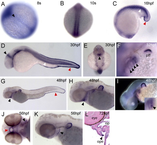
A: Expression of fras1 during development. fras1 expression is first noted in the axial midline 8 hr postfertilization (A, arrowhead). B-D: Transcripts are then detected in the developing somites (B, dorsal; C and D, white arrowheads). D,E: Expression is noted in the midbrain-hindbrain boundary (D, black arrowhead) toward the medial aspect of the future cerebellum (E, arrowhead). F: Transcripts are detected in the branchial arches specifically in the differentiating endodermal pouches (arrowheads). C,D,G: Strong expression is noted in the caudal fin fold shortly after its differentiation at the early tail bud stage (red arrowheads). F-I,K: Expression is detected in the pectoral fin fold (F) and this persists throughout the period investigated (G-I,K). G,H: By 48 hpf expression is noted in the oral ectoderm (black arrowheads) and in the posterior margin of the hyoid arch. I: By this stage, expression in the branchial arches is strongest in the pouches 4 and 5, in the most ventral aspects of these structures (inset arrowhead). J-L: Expression in the oral ectoderm persists at 56 hpf (J, red arrowhead) as does expression in the posterior ectodermal margin (PEM) at 56 hpf (J and K, black arrowheads) and 72 hpf (L, section, arrowhead). op, oropharynx; ope, operculum. EXPRESSION / LABELING:
|
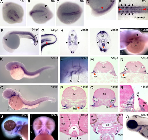
Expression of frem3 during development. frem3 is broadly expressed during embryonic development in zebrafish. A-C: Expression is first detected during early somitogenesis and, at the bud stage is detected in the lateral head mesoderm (A, anterior), somites (B, posterior; C, dorsal), and differentiating tail ectoderm (B, arrowhead). E,I: In newly differentiating caudal somites, expression is highest in the adaxial cells adjacent to the neural tube (black arrowheads). F,H: By 24 hpf expression is detected throughout the somitic musculature (F, lateral; H, in section, arrowhead). J: Expression is noted in the developing pectoral fin musculature (asterisk) which persists in the adjacent somites 1 and 2 (arrowhead). F-I: As with the other frem-related genes, expression is detected in the caudal fin folds. D,E,G,I: In addition to somitic expression, transcripts are observed in the hypochord (red arrowheads) as a single medial line of cells. K-R: During branchial arch development frem3 is expressed in the endodermal pouches (K-O; in section P,Q,R, red arrowheads). Expression is also noted in the otic placode (F, arrow) and by later developmental stages this expression is highest in the dorsolateral aspect of the otic vesicle (N,Q). S-V: Expression is initiated in the musculature of the developing head and in the hyoid posterior ectodermal margin and extending into the medial ectoderm (S-U, and in section, V). W: Expression in the operculum persists during later embryogenesis. nt, neural tube; nc, notochord; hb, hindbrain; bae, branchial arch endoderm; ov, otic vesicle; pff, pectoral fin fold; som, somite; pfm, pectoral fin muscle; op, oropharynx; ia, intermandibularis posterior muscle; ope, operculum. EXPRESSION / LABELING:
|
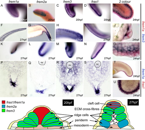
Expression of the frem and fras genes during median fin morphogenesis. Upper panel: Expression of the frem/fras genes in the developing fin fold. At 20 hours postfertilization (hpf), expression of frem1a and fras1 are detected in a midline stripe of cells (A,D), whereas frem3, and to a lesser extend frem2a, are expressed in two complementary cell populations cells (B,C). At 27 hpf, this pattern of complementary expression is continued, although frem2a is now expressed in both medial and lateral cell populations (F-I, dorsal/ventral; K-N, posterior. Dual in situ hybridization confirms complementary expression of frem3 and frem1a (E, lateral; J, section; O, lateral) and co-expression of frem1a and fras1 (T,U). Sections of embryos at 27 hpf confirm the observation made in whole embryos (P-S, caudal fold). Lower panel: We propose a model of complementary and overlapping gene expression, which corresponds to ridge and cleft cell populations first identified morphologically by Dane and Tucker,[1985]. nc, notochord. EXPRESSION / LABELING:
|

Unillustrated author statements EXPRESSION / LABELING:
|