
- Title
-
Bmp activity gradient regulates convergent extension during zebrafish gastrulation
- Authors
- Myers, D., Sepich, D., and Solnica-Krezel, L.
- Source
- Full text @ Dev. Biol.
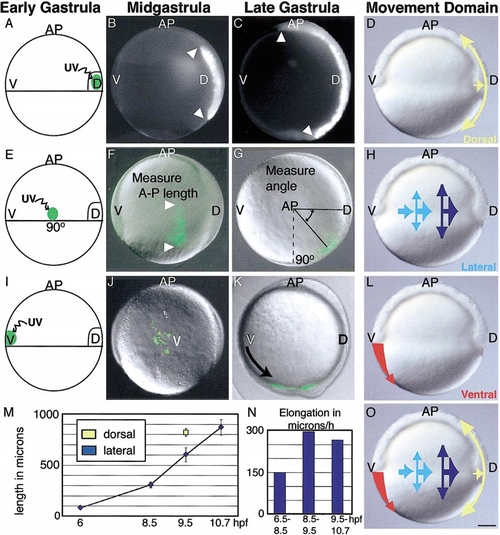
Domains of distinct convergence and extension movements in the zebrafish gastrula. The movements of groups of mesendodermal cells were examined in three regions: (A) dorsal, (E) lateral, and (I) ventral. Cells were labeled by photoactivation of a caged fluorescein dye. Resulting labeled cell arrays are illustrated at midgastrulation (8.5 hpf; B, F, J) and at the end of the gastrula period (9.5 hpf; C, G, K). (B, C, F) Elongation of dorsal and lateral cells groups was measured to monitor extension movements (M), and the rate of elongation of lateral cell arrays is shown in (N). (G) Dorsalward translocation of lateral cells was measured to monitor convergence. (I) Labeled cells of the ventral margin in WT gastrulae move underneath ectoderm and spread over the yolk (J), then into the tailbud region (K). From these and previous experiments (Sepich et al., 2000), three movement domains are inferred: (D) a dorsal region of strong extension and moderate convergence (yellow arrow), (H) a lateral domain of increasing convergence and extension (light blue to dark blue arrows), and (L) a ventral domain of inhibited convergence and extension (red arrow). This model is summarized in (O). Scale bar, 100 μm. |
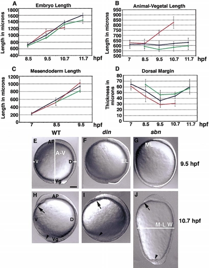
Increase and decrease in Bmp activity have distinct effects on CE of tissues and embryonic morphology. Graphs showing results of morphometric analysis of WT (blue), sbn (red), and din (green) mutant embryos: (A) embryo length, (B) animal–vegetal length, (C) dorsal mesendodermal length, and (D) thickness of dorsal margin. (E) Lateral view of a WT embryo at the end of the gastrula period (9.5 hpf) with animal pole (AP), vegetal pole (Vg), ventral (V), and dorsal (D) marked. The dorsal and ventral margin thickness and the animal–vegetal dimension are shown. (F) din embryo at 9.5 hpf; embryo length measure is shown. (G) sbn embryo at 9.5 hpf; mesendodermal length measure is indicated (ML). (H) WT, (I) din, and (J) sbn embryos at the beginning of segmentation; mediolateral embryo width is indicated (M-LW; 10.7 hpf). Arrows denote anterior tip of mesendoderm, and arrowheads mark tail region (H–J). A–V, animal–vegetal length; EL, embryo length; ML, mesendoderm length; M-LW, mediolateral width. Scale bar, 100 μm. |
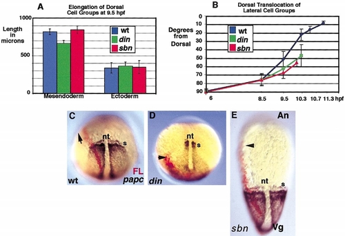
Elongation of dorsal mesendodermal cell populations, dorsal translocation, and fate-mapping analyses of lateral cell groups in WT, din, and sbn mutants. (A) Elongation of labeled mesendodermal cell populations is reduced in din mutant gastrulae by the end of the gastrula period. (B) Assaying dorsal translocation of labeled lateral marginal cells indicates that convergence movements in both sbn and din mutants become impaired after midgastrulation. Fate-map analysis of lateral marginal cells in (C) WT, (D) din, and (E) sbn embryos at early segmentation; the resulting labeled lateral cell array is seen in red (FL). The anterior boundary of the labeled cell array is marked by an arrowhead; in WT, the array extends around the embryo into the head mesoderm and its anterior boundary is not visible, denoted by an arrow. paraxial protocadherin (papc) expression is shown in brown. s, somites; n, notochord. |
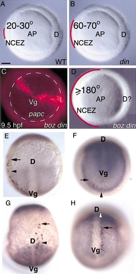
The NCEZ is specified by high levels of Bmp activity. (A) In WT, the NCEZ comprises 20–30° of the ventral margin. (B) The dorsally expanded Bmp activity in moderately ventralized din mutants leads to an enlargement of the NCEZ to a 60–70° arc of the ventral margin. (C) Further dorsal expansion of Bmp activity in boz din double mutants results in a dramatic NCEZ enlargement. Shown is a vegetal view of a boz din embryo, with a broken line marking the enlarged vegetal papc expression domain. Labeled cells moved into this vegetal region without translocating dorsally away from their original point of labeling. Furthermore, they fail to extend beyond the papc expression domain. (D) Cells from at least a 180° arc of the ventral margin constitute NCEZ in boz din mutants. (E) Dorsal view of a host embryo at the end of the gastrula period with transplanted cells (in brown) from a nonventralized host. At the beginning of gastrulation, the transplanted cells were in the ventral lateral region of the margin. (F) Cells that were transplanted from a strongly ventralized donor to the same ventral lateral region of the gastrula have moved into the tailbud region, shown in this vegetal view. (G) Dorsal view of cells transplanted from a nonventralized donor near the dorsal shield at the beginning of the gastrula period. They form arrays extending outside of the papc expression domain. (H) Dorsovegetal view of a host embryo with cells from a ventralized donor that were transplanted near the dorsal shield and have now moved into the posterior trunk and tailbud region. |
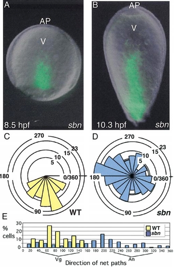
High levels of Bmp activity are necessary for NCEZ and promote vegetal migration of mesendodermal cells. (A) Ventral view of a labeled sbn embryo at midgastrulation; note the extended cell array. (B) sbn embryo at early segmentation showing a significantly elongated labeled cell array. (C) Ventral cells migrate vegetally in WT from midto late gastrulation. The direction of their net paths was determined from three time-lapse recordings from 8.2 to 8.8 hpf (75–85% epiboly). Rose diagrams are arranged to represent the number of ventral cells taking these net paths, with all paths starting at a common origin; vegetal is represented as 90° and animal direction as 270°. (D) Ventral mesodermal cells in sbn migrate along net paths oriented in all directions. (E) The same data are represented as the percentage of cells taking each net path, in order to demonstrate the strong vegetal bias in WT and slight lateral bias in sbn. Scale bar, 100 μm. |
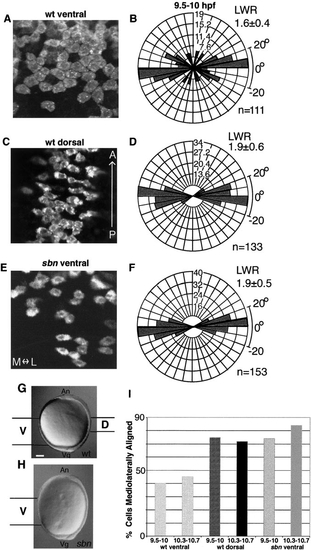
Cells at low levels of Bmp activity are mediolaterally aligned and elongated. Confocal microscope images of DiI-labeled cells from (A) WT ventral, (C) WT dorsal, and (E) sbn ventral regions (9.5–10.3 hpf). (B, D) Rose diagrams of cell orientations from WT and (F) sbn embryos at 9.5–10 hpf. The mediolateral axis corresponds to the horizontal plane, while the anteroposterior axis aligns vertically. Cell number scale is indicated on each diagram. (G, H) An overview of the locations of cells analyzed in WT and sbn embryos. (I) Graph shows percentage of analyzed cells, which are mediolaterally aligned, i.e., orient the long axis ±20° with respect to the mediolateral axis. Hatched bars indicate late gastrula stages (9.5–10 hpf); solid bars are early segmentation stages (10.3–10.7 hpf). n, number of cells analyzed; LWR, length/width ratio. Scale bar, 100 μm. |
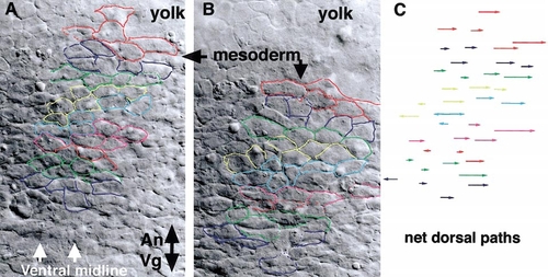
Dorsally biased mediolateral intercalation in ventral regions of sbn mutants. Time-lapse Nomarski image recordings were made of ventral mesodermal cells in sbn embryos beginning shortly after the end of the gastrula period (9.7 hpf) until the beginning of segmentation (10.3 hpf). (A) Initial image showing mesodermal cells overlaying the yolk, vegetal pole to the bottom. Cells that could be identified throughout the recording are outlined in color. (B) After 40 min, the analyzed cell population moved vegetally, with respect to the yolk. Individual cells intercalated, dorsally broadening the population in a mediolateral direction. The corresponding net path of each cell is shown in (C). |
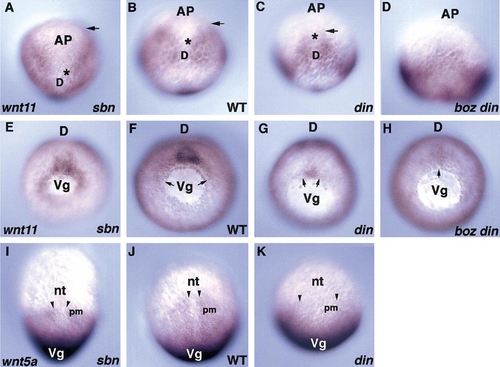
wnt11 (slb) (A–H) and wnt5a (ppt) (I–K) expression patterns during late gastrulation (9–9.2 hpf). (A–H) Asterisks denote dorsal boundary of anterior wnt11 expression domain, while arrows indicate ventral boundary. (A) Dorsoanterior view of sbn gastrulae showing ventrally expanded expression of wnt11. Dorsal views of (B) WT, (C) din, and (D) boz din embryos showing progressive reduction of the anterior wnt11 expression domain in the ventralized mutants. Vegetal views of (E) sbn, (F) WT, (G) din, and (H) boz din embryos showing expansion in dorsalized and reduction in ventralized mutants of the wnt11 expression domain near the blastopore. Lateral boundaries of the blastopore expression are marked by arrows. Dorsal views of (I) sbn, (J) WT, and (K) din embryos showing wnt5a expression in presomitic mesoderm in sbn and WT and its loss in ventralized din mutants. Dorsal limits of expression are indicated by arrowheads. AP, animal pole; D, dorsal; Vg, vegetal pole; nt, notochord; pm, presomitic mesoderm. |
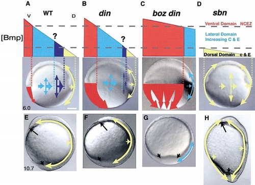
Model: Bmp activity gradient specifies convergence and extension movement domains. Thresholds of Bmp activity (dashed gray lines) specify three CE movement domains. (A) In WT, high ventral Bmp levels specify NCEZ (red). An intermediate level of Bmp activity specifies increasing convergence and extension, where cells initially converge and extend slowly (light blue) and then at a faster speed (dark blue). The transition point from slow to fast convergence is unclear (question mark; light blue to dark blue). In contrast, low levels of Bmp activity specify generous extension with limited convergence (yellow). In DV patterning mutants, these movement domains are altered according to changes in the Bmp activity gradient. In ventralized (B) din and (C) boz din, the NCEZ is enlarged with increased Bmp activity, while convergence and extension are reduced in lateral regions. The domain of increasing lateral convergence and dorsal convergent extension are also reduced in din and may be absent in boz din double mutants. (D) However, in sbn, low levels of Bmp activity around the circumference of the embryo promote strong extension and low convergence in all regions. The distribution and degree of these morphogenetic movements shapes the early segmentation embryo. (E) In WT, NCEZ cells move to the tailbud, while lateral movements sweep cells toward the dorsal domain, where CE drives the elongation of the axis (yellow). (F, G) In din and boz din, due to reduced lateral convergence and extension, fewer cells contribute to the anterior structures, while expanded NCEZ creates enlarged tailbud, increasing posterior structures at the expense of head and trunk. (H) In sbn, circumferential CE movements elongate the An-Vg axis, and may eventually impair the extension of the embryonic axis. Anterior end of head (long arrow); posterior end of axis, tailbud (short arrow). Scale bar, 100 μm. |
Reprinted from Developmental Biology, 243(1), Myers, D., Sepich, D., and Solnica-Krezel, L., Bmp activity gradient regulates convergent extension during zebrafish gastrulation, 81-98, Copyright (2002) with permission from Elsevier. Full text @ Dev. Biol.