
Fig. 1
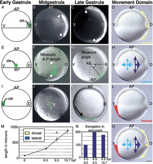
Domains of distinct convergence and extension movements in the zebrafish gastrula. The movements of groups of mesendodermal cells were examined in three regions: (A) dorsal, (E) lateral, and (I) ventral. Cells were labeled by photoactivation of a caged fluorescein dye. Resulting labeled cell arrays are illustrated at midgastrulation (8.5 hpf; B, F, J) and at the end of the gastrula period (9.5 hpf; C, G, K). (B, C, F) Elongation of dorsal and lateral cells groups was measured to monitor extension movements (M), and the rate of elongation of lateral cell arrays is shown in (N). (G) Dorsalward translocation of lateral cells was measured to monitor convergence. (I) Labeled cells of the ventral margin in WT gastrulae move underneath ectoderm and spread over the yolk (J), then into the tailbud region (K). From these and previous experiments (Sepich et al., 2000), three movement domains are inferred: (D) a dorsal region of strong extension and moderate convergence (yellow arrow), (H) a lateral domain of increasing convergence and extension (light blue to dark blue arrows), and (L) a ventral domain of inhibited convergence and extension (red arrow). This model is summarized in (O). Scale bar, 100 μm. |
Reprinted from Developmental Biology, 243(1), Myers, D., Sepich, D., and Solnica-Krezel, L., Bmp activity gradient regulates convergent extension during zebrafish gastrulation, 81-98, Copyright (2002) with permission from Elsevier. Full text @ Dev. Biol.