
Fig. 7
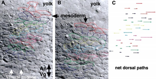
Dorsally biased mediolateral intercalation in ventral regions of sbn mutants. Time-lapse Nomarski image recordings were made of ventral mesodermal cells in sbn embryos beginning shortly after the end of the gastrula period (9.7 hpf) until the beginning of segmentation (10.3 hpf). (A) Initial image showing mesodermal cells overlaying the yolk, vegetal pole to the bottom. Cells that could be identified throughout the recording are outlined in color. (B) After 40 min, the analyzed cell population moved vegetally, with respect to the yolk. Individual cells intercalated, dorsally broadening the population in a mediolateral direction. The corresponding net path of each cell is shown in (C). |
Reprinted from Developmental Biology, 243(1), Myers, D., Sepich, D., and Solnica-Krezel, L., Bmp activity gradient regulates convergent extension during zebrafish gastrulation, 81-98, Copyright (2002) with permission from Elsevier. Full text @ Dev. Biol.