
|
Fig. 4
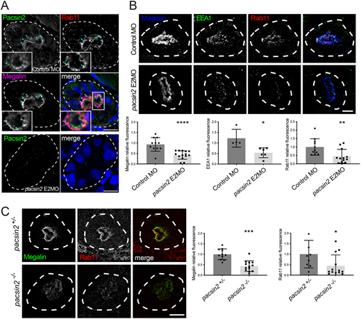
Loss of Pacsin2 causes reduced abundance of megalin and endocytic markers in the proximal tubule. (A) Top, transverse cryosections of proximal tubule were immunolabeled for Pacsin2, Rab11 and megalin in 3 dpf wild-type larvae. Insets depict an enlarged view of the boxed region, rotated 90˚ clockwise. Blue arrows indicate regions of colocalisation between Pacsin2, Rab11 and megalin. Bottom, 3 dpf pacsin2 E2MO morphant larvae labelled with anti-Pacsin2 antibody. (B) Top, transverse cryosections through the pronephros of 3 dpf control morphant or pacsin2 E2MO morphant larvae immunolabeled for megalin, EEA1 and Rab11. Bottom, quantification of relative signal intensity of megalin, EEA1 and Rab11, respectively, between control and pacsin2 morphants. n=5-12 for control morphants and 6-14 for pacsin2 morphants. Unpaired t-test, * P<0.05, ** P<0.01, **** P<0.0001. Error bars=s.d. (C) Left, transverse sections through the proximal tubule of 4 dpf pacsin2+/− or pacsin2−/− larvae immunolabelled for megalin and Rab11. Right, quantification of relative signal intensity of megalin and Rab11, respectively, between pacsin2+/− and pacsin2−/− larvae. n=11 and 13 for megalin, and 9 and 13 for Rab11 for pacsin2+/− and pacsin2−/−, respectively. Unpaired t-test, *P<0.05 *** P<0.001. Error bars=s.d. Dashed white lines indicate the outer margin of the pronephros. Scale bars: 10 µm.