IMAGE

Fig. 2

ID
ZDB-IMAGE-200406-149
Genes
Antibodies
Source
Figures for Zhao et al., 2020
Image
Figure Caption

Fig. 2

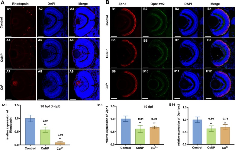
Protein levels of retinal photoreceptor in copper stressed embryos. A Immunostaining of Rodopsin (labeling retinal rod cells) (A1-A9). B Double immunostaining of ZPR-1 (labeling retinal cone cells) and Opn1sw2 (blue opsin, labeling retinal rod cells) in embryos at 10 dpf. A10, B13, B14, Quantification of protein level in each sample. Scale bars: A1-A9 and B1-B12, 50 μm; **, P < 0.01; *, P < 0.05

Figure Data
Acknowledgments
This image is the copyrighted work of the attributed author or publisher, and ZFIN has permission only to display this image to its users. Additional permissions should be obtained from the applicable author or publisher of the image. Full text @ Cell Commun. Signal.