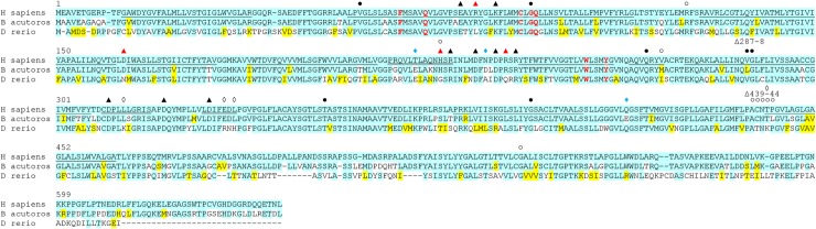
IMAGE

Fig 1

ID
ZDB-IMAGE-200229-20
Source
Figures for Concilio et al., 2020
Image
Figure Caption

Fig 1 Amino acid sequence alignment between the NIS proteins of <italic>H</italic>. <italic>sapiens</italic> (human), <italic>B</italic>. <italic>acutorostrata scammoni</italic> (minke whale), and <italic>D</italic>. <italic>rerio</italic> (zebrafish).

Cyan highlighting indicates absolute conservation to human NIS. Yellow indicates similar residue to human NIS. Underline indicates putative transmembrane domain in human NIS, only TM1-12 are indicated. Closed circles indicate site of a mutation known to cause a transport defect in humans [22]. Open circles indicate site of a mutation known to cause membrane trafficking defect in humans [22]. Black triangles indicate a charged residue where mutation to alanine significantly reduces iodide uptake in human NIS [71]. Red triangles indicate a charged residue where mutation to alanine significantly reduces iodide uptake in human NIS and this residue is not charged in zebrafish NIS [71]. Open diamonds indicate additional positively charged residues in zebrafish NIS. Blue diamonds indicate additional negatively charged residues in minke whale NIS. Bold red lettering indicates residue reported to be involved in stoichiometry control and translocation dynamics [5253]. Numbering follows human NIS.

Acknowledgments
This image is the copyrighted work of the attributed author or publisher, and ZFIN has permission only to display this image to its users. Additional permissions should be obtained from the applicable author or publisher of the image. Full text @ PLoS One