
|
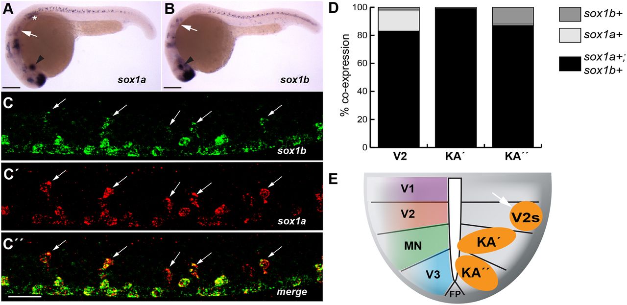
Fig. 1
sox1a and sox1b are co-expressed in the zebrafish spinal cord. (A,B) Embryos at 24 hpf hybridized to sox1a (A) and sox1b (B) probes. Both genes are expressed in the forebrain, hindbrain and lens (arrowheads), in the otic vesicle (arrows), and in cells along the spinal cord. In addition, sox1a is strongly expressed in the lateral line primordium (asterisk). (C-C″) Fluorescence in situ hybridization (FISH) for sox1a and sox1b mRNA at 24 hpf shows co-expression in V2 (white arrows) and KA neurons. (D) Cells were counted over the yolk extension in a five-somite-long segment. Data are mean±s.d. In the V2 domain, 83±2% of cells (n=103) co-express sox1a and sox1b mRNA, 15±3% of cells express only sox1a, and 2±1% of cells express only sox1b. Similar counts were obtained for KA′ (sox1a/b, 99±4%; sox1a, 0%; sox1b, 1±0%, n=86) or KA″(sox1a/b, 87±3%; sox1a, 1±0%; sox1b, 12±3%, n=115). Six embryos from two independent experiments were counted. (E) Ventral spinal cord domains V1 to V3 with the locations of sox1a+ and sox1b+ KA′ and KA″ neurons (orange circles), and neurons in the V2 domain (V2s, arrow) indicated. Embryos are at 24 hpf. Dorsal is upwards; anterior is leftwards. Scale bars: 200 μm in A,B; 25 μm in C-C″.