
|
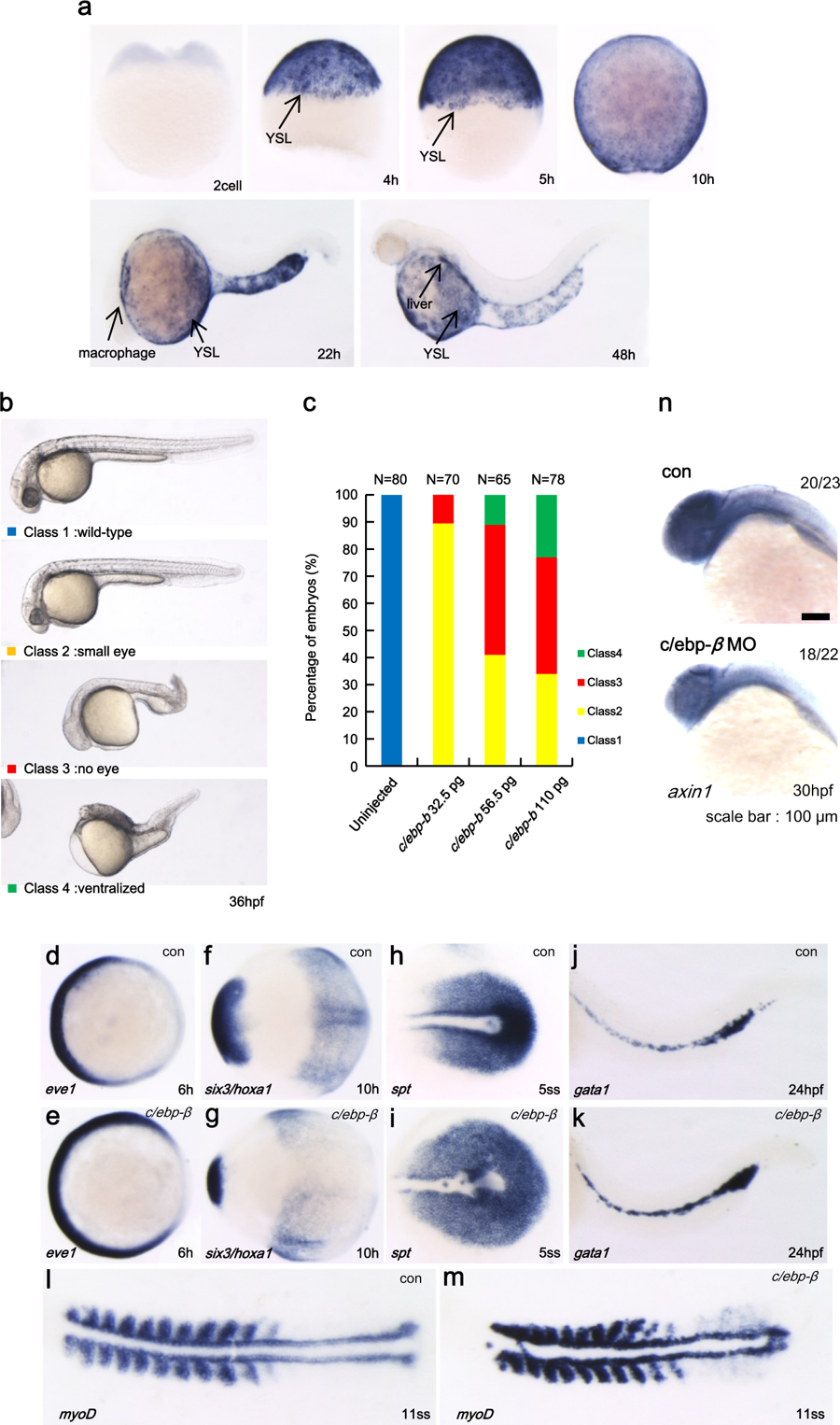
Fig. 5
a Spatial and temporal expression of c/ebp-β in developing zebrafish embryos determined by whole-mount in situ hybridization. b Injection of zebrafish c/ebp-β mRNA induced ventralized phenotype at 36 hpf embryos. c Percentage of embryos displaying specific phenotypes following c/ebp-β mRNA injection. d–m Expression of marker genes in c/ebp-β injected embryos. eve1, a ventral marker (d, e); six3, anterior neural marker; hoxa1, posterior neural marker at the tail bud stage (f, g); spt, posterior mesoderm marker (h, i) in intermediate cell mass (ICM) in the 5-somite stage embryo; gata1 expression in ICM (j, k); myoD expression in the 11-somite stage embryo (l, m). Embryos are shown in a dorsal view with anterior to the left. cont control mRNA, 5ss 5-somatic stage, 11ss 11-somatic stage. n The expression of axin1 was examined by whole-mount in situ hybridization analysis after c/ebp-β MO injection. con control MO