
|
Fig. 6
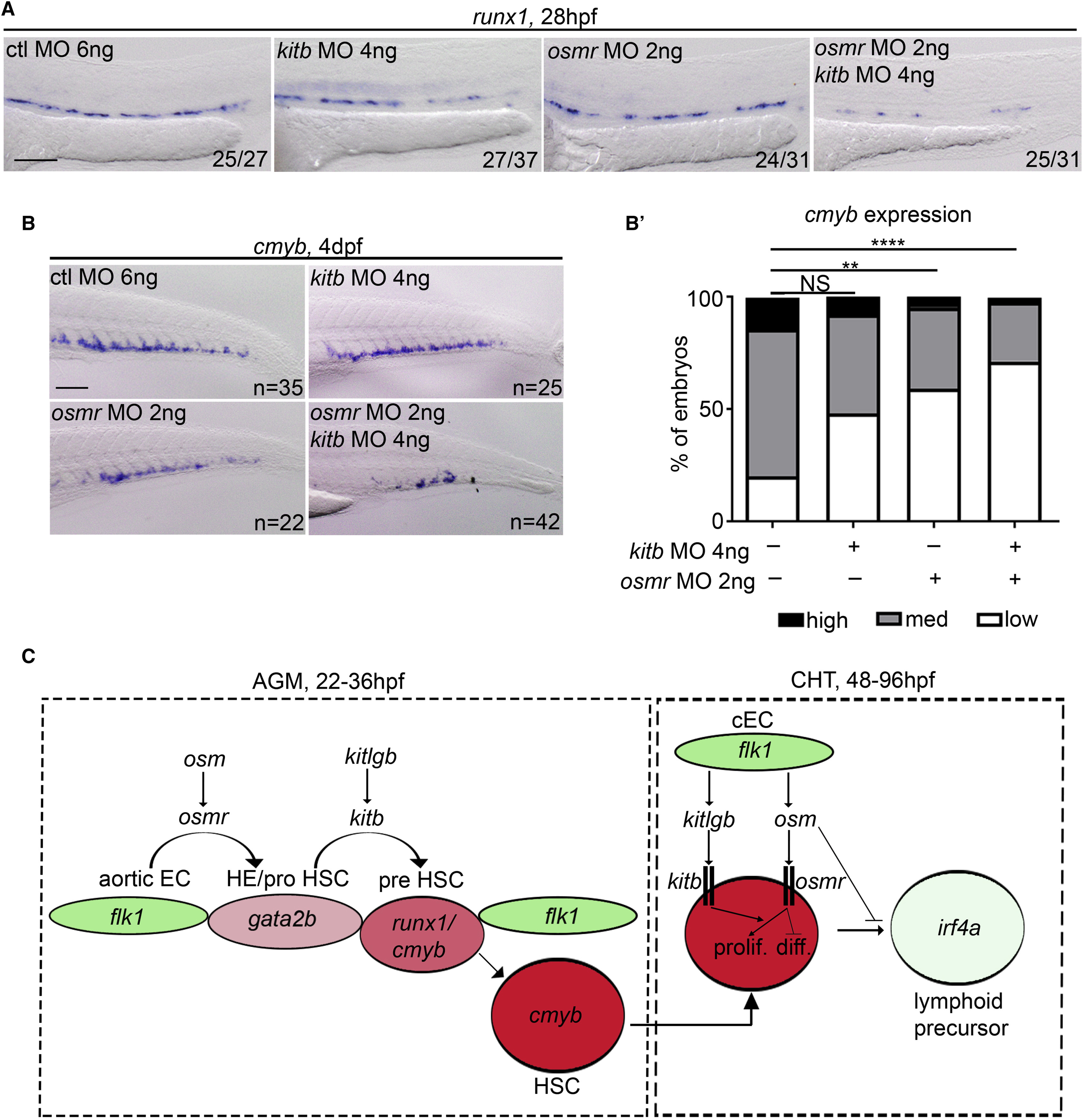
Inhibition of osmr and kitb Signaling Synergistically Inhibits HSC Specification
(A) runx1 (28 hpf) ISH following control MO, osmr MO, or kitb MO injection (separately and together) at subliminal doses (half doses). ctl, control.
(B) cmyb ISH at 4 dpf following control MO, osmr MO, or kitb MO injection (separately and together) at subliminal doses (half doses). (B′) Quantification of the results observed in panel (B). High cmybsignal (black), medium cmyb signal (grey) and low cmyb signal (white). Analysis was generated by Fisher’s exact test. NI versus kitb-MO; p = 0.082, NI versus osmr-MO; p = 0.0089, NI versus kitb+osmr-MOs; p = 0.0000009.
(C) Summary. Aortic ECs transition to “HE/pro HSC” by expressing gata2b, a process that requires osm/osmr signaling. HSCs then begin to bud from the aorta and begin to express runx1 to become “pre HSCs”, a process that requires kitlgb/kitb signaling. HSC then become “HSCs” and express cmyb. HSCs migrate to the CHT and can either proliferate (prolif.) or differentiate (diff.) in response to cytokines expressed by caudal ECs (cECs). Some will differentiate to lymphoid lineages. Proliferation of HSCs is enhanced by osm and differentiation to lymphoid lineages is inhibited. HSC proliferation is also synergistically enhanced by both kitlgb and osm signaling through their receptors. ∗∗∗∗p < 0.0001; ∗∗p < 0.01. All scale bars, 100 μm. HE, hemogenic endothelium.