
Fig. 1
- ID
- ZDB-IMAGE-180816-1
- Genes
- Publication
- Davis et al., 2018 - ETS transcription factor Etsrp / Etv2 is required for lymphangiogenesis and directly regulates vegfr3 / flt4 expression
- All Figures
- Figures for Davis et al., 2018
|
Fig. 1
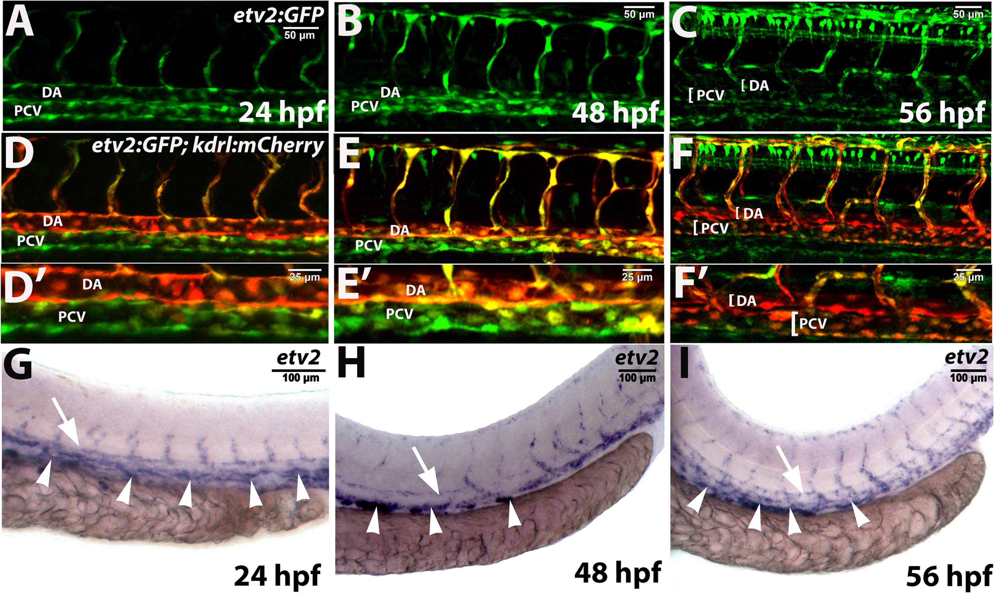
Etv2 expression in wild type embryos and larvae as analyzed by the transgenic reporter expression and in situ hybridization. (A-F) Confocal images of Tg(−2.3 kb etv2:GFP; kdrl:mCherry) reporter line at 24–56 hpf. Note that etv2:GFP expression is downregulated in the dorsal aorta (DA) at 48–56 hpf but is present in multiple cells in the posterior cardinal vein (PCV). (A-C) and (D-F) show the same embryos and larvae in green and overlaid green and red channels. (D′-F′) are higher magnification images of the DA and PCV from embryos and larvae in (D-F). Non-specific expression of etv2:GFP in neural cells is also apparent in (C,F). (G-I) In situ hybridization analysis of etv2 expression at 24–56 hpf. Note the enriched etv2 expression in the PCV (arrowheads) as compared to the DA (arrows) at 48–56 hpf. Each assay has been repeated at least twice with over 10 embryos or larvae analyzed in each group.
Reprinted from Developmental Biology, 440(1), Davis, J.A., Koenig, A.L., Lubert, A., Chestnut, B., Liu, F., Desai, S.P., Winkler, T., Pociute, K., Choi, K., Sumanas, S., ETS transcription factor Etsrp / Etv2 is required for lymphangiogenesis and directly regulates vegfr3 / flt4 expression, 40-52, Copyright (2018) with permission from Elsevier. Full text @ Dev. Biol.