
|
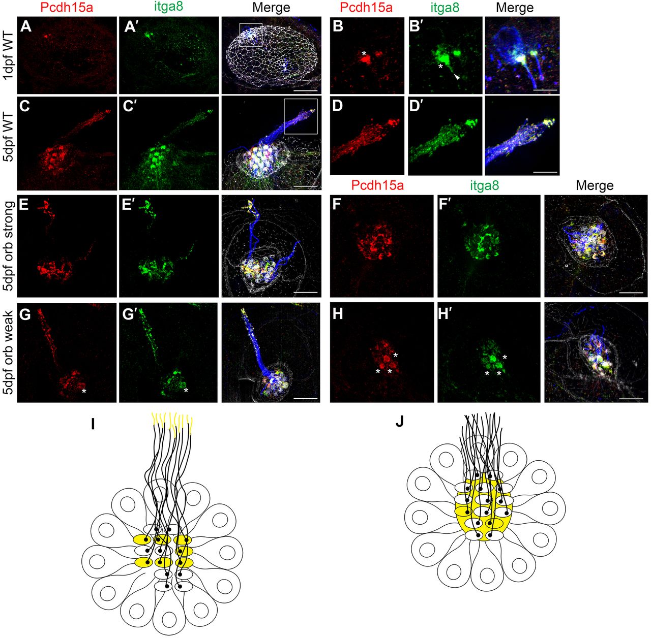
Fig. 3
Itga8 and Pcdh15a localize at the apical aspect of hair cells. Representative images of embryos/larvae immunostained for Pcdh15a (red), Itga8 (green) and acetylated tubulin (blue). Samples were counterstained with phalloidin (gray). (A–B′) Confocal microscopy analyses of a 1 dpf zebrafish inner ear (dorsal view, anterior to the left). (B,B′) Magnification of boxed area in A,A′. (C–H′) SR-SIM of 5 dpf neuromasts. (C–D′) WT. (D,D′) Magnification of boxed area in C,C′. (E–F′) orbiter strong; (G–H′) orbiter weak. Asterisks denote hair bundle immunostaining. The arrowhead in panel B′ denotes cilia immunostaining. (I,J) Cartoons of neuromasts (top views) showing colocalization (yellow) of Itga8 and Pcdh15a. In WT animals (I), both proteins colocalize at the hair bundle in a sub-set of cells and towards the tip of the kinocilia. In mutant animals (J), there is some colocalization at the hair cell bundle but also around it (cuticular plate). Scale bars: 25 µm (A), 8 µm (B), 4 μm (D), 7 µm (C,E–H). Three independent experiments were performed.