
|
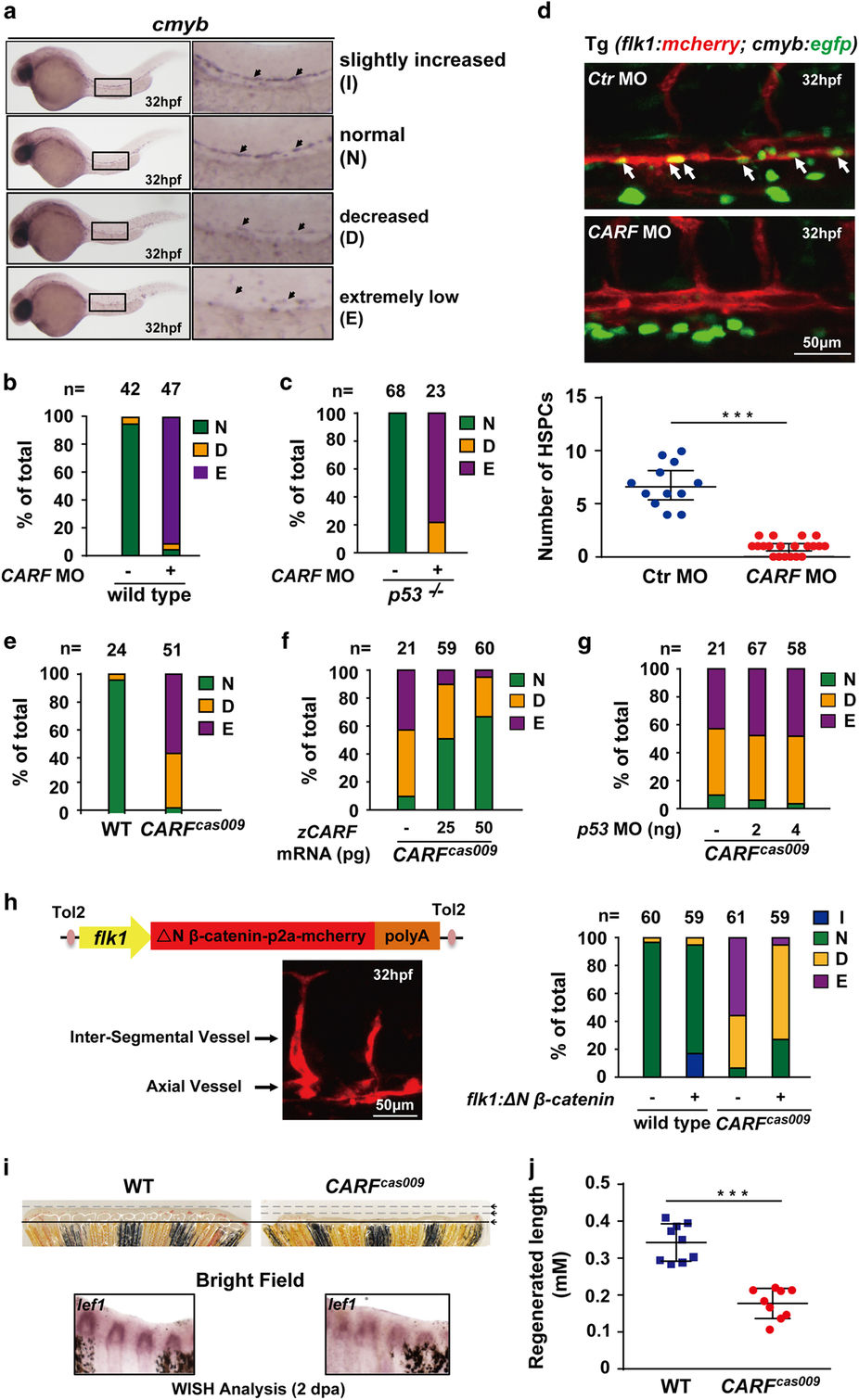
Fig. 5
Loss of CARF attenuates HSPC formation and caudal fin regeneration. (a–d) Knockdown CARF limits HSPC formation. Zebrafish embryos at 32 hpf were fixed for in situ hybridization with cmyb probe, and then classified into three categories: (a) Representative image for slightly increased (I), normal (N), decreased (D) or extremely low (E) level of c-myb WISH analysis. Knockdown CARF dramatically inhibits HSPC formation (b) which could not be rescued via p53 MO co-injection (c) and further validated by the live image of HSPC budding events in AGM of zebrafish Tg(flk1:mcherry;cmyb:egfp) line (d). (e–g) CARFcas009 mutant exhibits reduced HSPC formation via a p53-independent manner. Injection of zebrafish CARF (zCARF) mRNA (f) but not p53 MO (g) restores decreased cmyb expression in CARFcas009 mutants (e). (h) Schematic diagram of Tol2 transposase-mediated transient transgenesis of endothelial-specific promoter (flk1)-derived expression of constitutive activated β-catenin (ΔN β-catenin) and mCherry chimera protein in zebrafish embryos, which rescues hematopoietic defects in CARFcas009 mutants while slightly increases HSC formation in wildtype zebrafish. (i, j) Delayed fin regeneration of CARFcas009 mutants. Adult CARFcas009 zebrafish were executed caudal fin amputation and then cultured for regeneration. The regenerated sections were cut down at 2 dpa for either WISH analysis with lef1 probes or length measurement.