
|
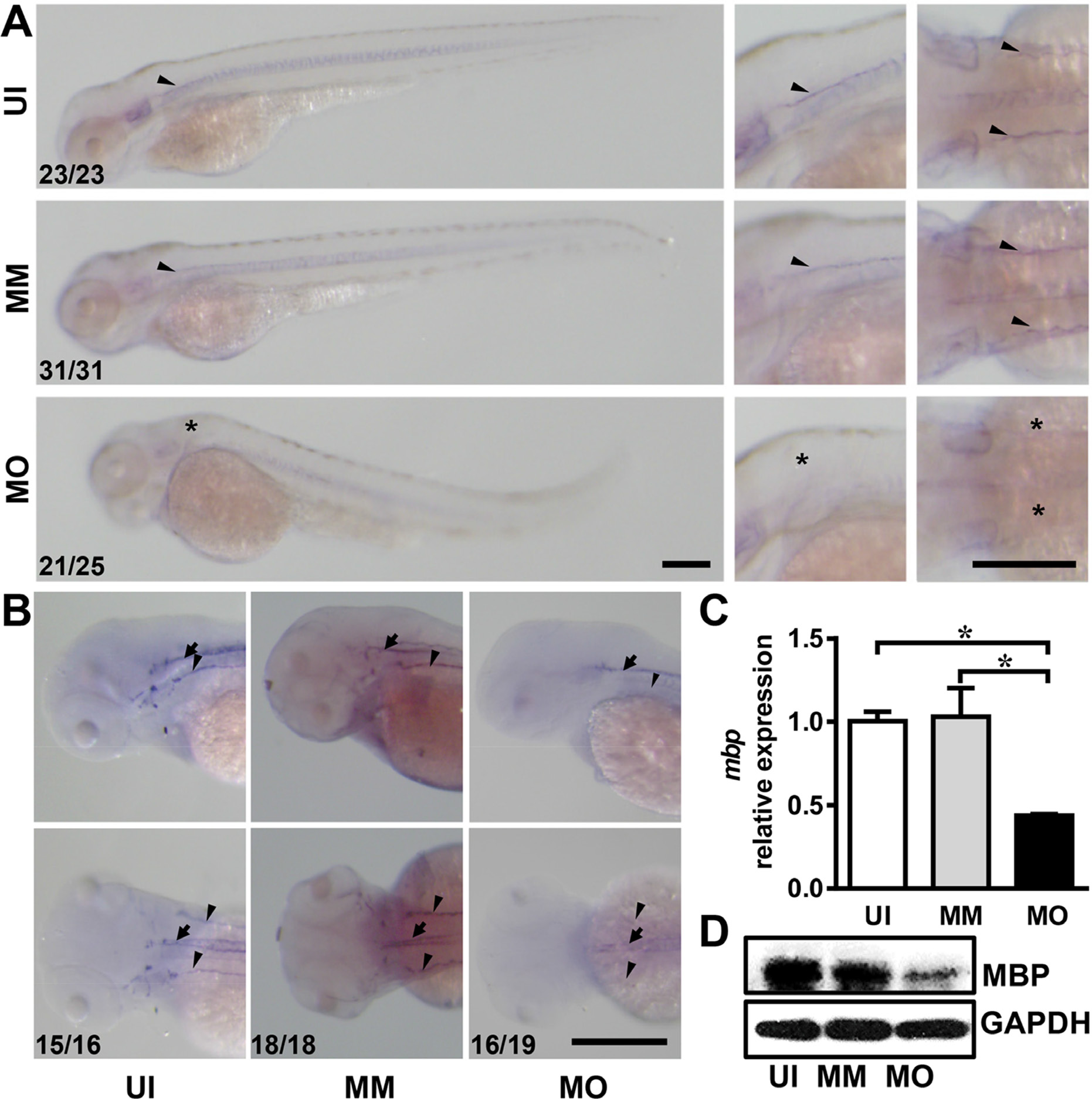
Fig. 4
IL-7R knockdown delays myelination in the nervous system. (A) The images of mbp mRNA expression in embryos from uninjected (UI), mismatch control (MM) and IL-7R morphant (MO) at 3 dpf. The second and third lines are magnified images in lateral view and dorsal view. Note that the mbp-expressing cells in IL-7R morphant are absent at 3 dpf (asterisks). (B) The images of mbp expression in the spinal cord (arrows) and lateral line nerves (arrowheads) of the larvae from the UI, MM and MO groups at 4dpf. The mbp positive cells distribute along the lateral line nerves and the spinal cord in UI and MM larvae. In MO larva, the mbp signals are only detected along the spinal cord. (C) The relative expression of mbp mRNA expression among three groups using qRT-PCR at 4 dpf (n = 3 in each group). Note that the expression level in the MO group is decreased compared to the UI and MM groups (ANOVA, *P < 0.05). (D) The expression of MBP protein by western blotting at 4 dpf. Scale bar: A. and B., 200 μm.