
|
Fig. S2
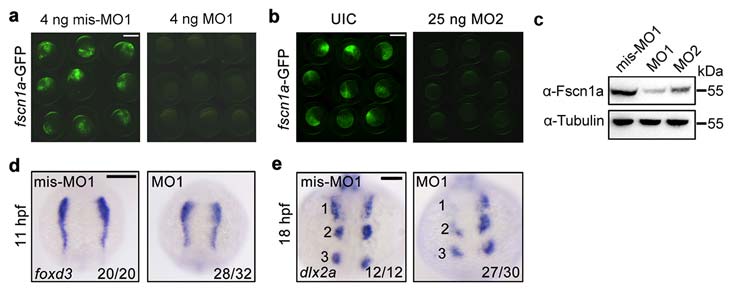
fscn1a morphants exhibit NC cell migration defects in the first stream.
(a-b) Effectiveness of fscn1a MO1 and MO2. Embryos co-injected with 50 pg fscn1a-GFP plasmid DNA and indicated MOs. Green fluorescence was detected at the shield stage. fscn1a MO1 (a) or MO2 (b) injected embryos showed a noticeable decrease in green fluorescence when compared to control embryos. Scale bar, 400 µm. (c) The expression of fscn1a protein was examined by Western blots in shield-stage embryos injected with fscn1a MO1 (4 ng) or MO2 (25 ng). The expression of Tubulin was detected as loading control. (d-e) Embryos injected with mis-MO1 (4 ng) or fscn1a MO1 (4 ng) at the one-cell stage and harvested at indicated stages for in situ hybridization with foxd3 (d) and dlx2a (e) probes. Dorsal views with anterior to the top. Numbers were labeled to show the three streams of pharyngeal NC cells. Scale bar, 200 µm in panel d and 100 µm in panel e.