
|
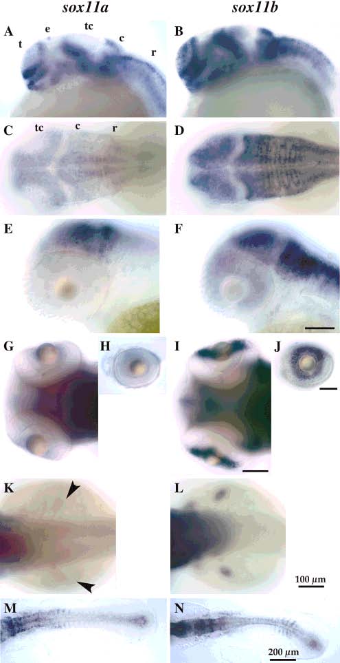
Fig. 4
Sox11a and sox11b genes are expressed in overlapping and distinct regions of the embryo. A,B; E,F; H,J: Side views, anterior to the left. C,D; G,I; K-N: Dorsal views, anterior to the left. Scale bar = 100 µm unless otherwise indicated. t, telencephalon; e, epiphysis; tc, tectum; c, cerebellum; r, rhombencephalon. A,B: 24 hr embryos, sox11a is expressed in epiphysis (e), but not the tectum (tc); sox11b is expressed in the tectum, but not the epiphysis. C,D: 48 hr embryos, sox11b but not sox11a is expressed in the anterior lateral tectum at 48 hr. E,F: 48 hr embryos focused on the eye. G-J: 80 hr embryos, only low levels of sox11a are found in the eye at 80 hr, whereas sox11b is strongly expressed. K,L: 48 hr embryos, sox11b but not sox11a is expressed in the prospective fin buds at 48 hr. M,N: Flatmount of 14-somite stage embryos reveal differential expression of sox11a in the anterior somites and sox11b in the posterior somites.