
Image
|
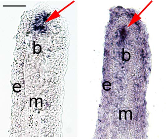
Figure Caption
Fig. 2
In situ hybridization of esco2 in fin cryosections. Left: Cryosection from a whole-mount WT-5 dpa fin treated for in situ hybridization showing tissue specific expression of esco2 mRNA within the blastema compartment. Right: In situ hybridization was completed on a WT-5 dpa fin cryosection showing a similar pattern of esco2 expression localized specifically at the blastemal region. Arrows indicate esco2 expression at blastema. B, blastemal; e, epidermis; m, mesenchyme. Scale bar = 50 µm.
Figure Data
Acknowledgments
This image is the copyrighted work of the attributed author or publisher, and
ZFIN has permission only to display this image to its users.
Additional permissions should be obtained from the applicable author or publisher of the image.
Full text @ Dev. Dyn.