
|
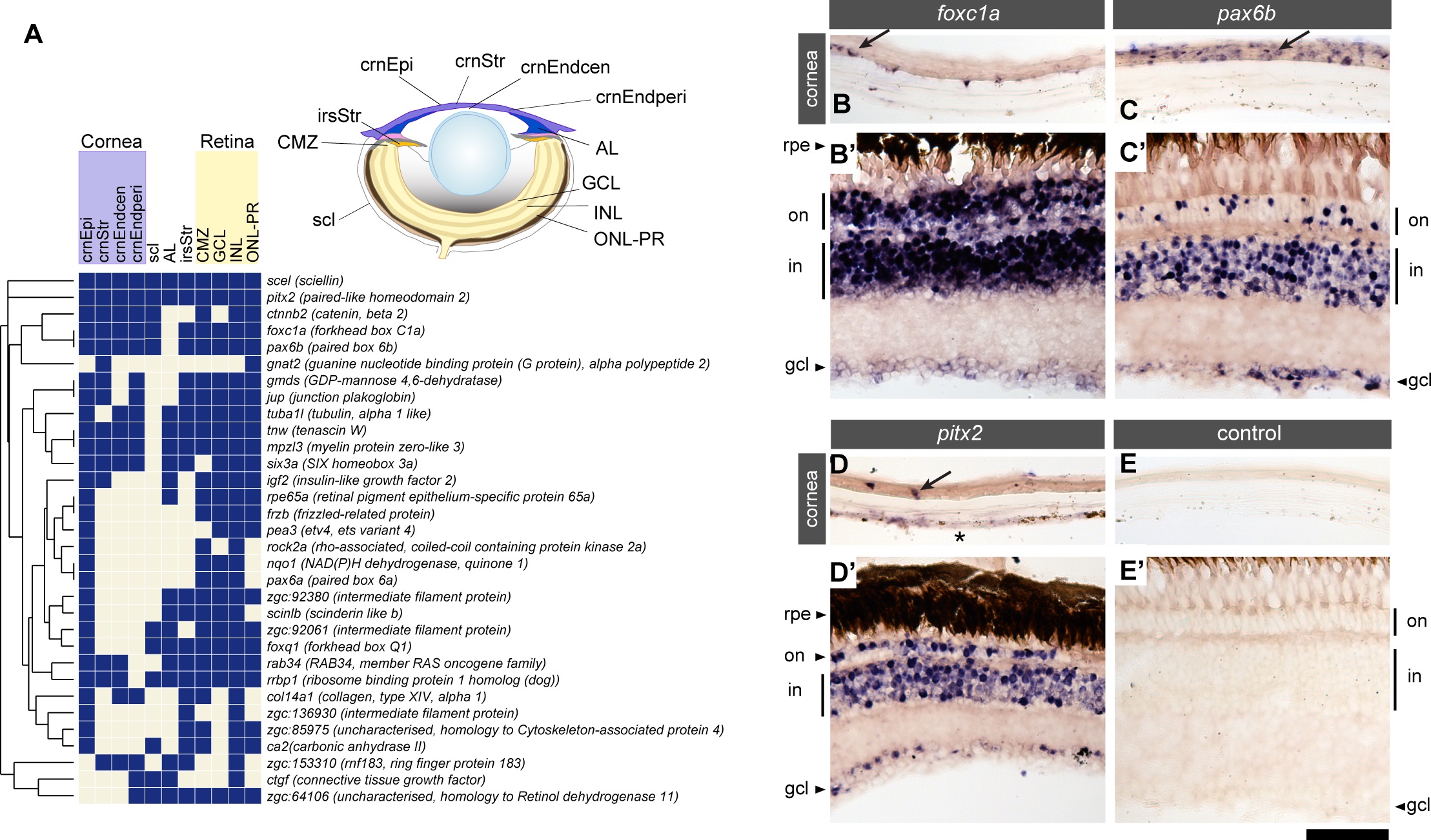
Fig. 3
Genes expressed in the cornea are also expressed in the retina of the adult eye.
(A) Analysis of genes and their expression in structures of the adult zebrafish eye. The 32 genes examined are listed along the vertical axis. The examined anatomical structures are shown in the schematic and listed on the horizontal axis. Anatomical structures examined are inner nuclear layer of the retina (INL), iris stroma (irsStr), outer nuclear layer and outer plexiform layer (ONL-PR), ganglion cell layer (GCL), ciliary marginal zone of the retina (CMZ), corneal epithelium (crnEpi), annular ligament (AL), sclera (scl), corneal stroma (crnStr), peripheral region of the corneal endothelium (crnEndperi) and central region of the corneal endothelium (crnEndcen). (B-E): Examples of genes expressed in the cornea and the retina. in situ gene expression patterns of foxc1a (B, B′), pax6b (C, C′) and pitx2 (D, D′) are shown in the adult cornea (B-D) and retina (B′-D′), with tissue processed without antisense mRNA probe as a negative control (E, E′). Genes expressed in either the corneal epithelium (arrows) or the corneal endothelium (asterisk) are also expressed in the retina. rpe: retinal pigment epithelium; on: outer nuclear layer; in: inner nuclear layer; gcl: ganglion cell layer. Scale bar: 50 µm.