
|
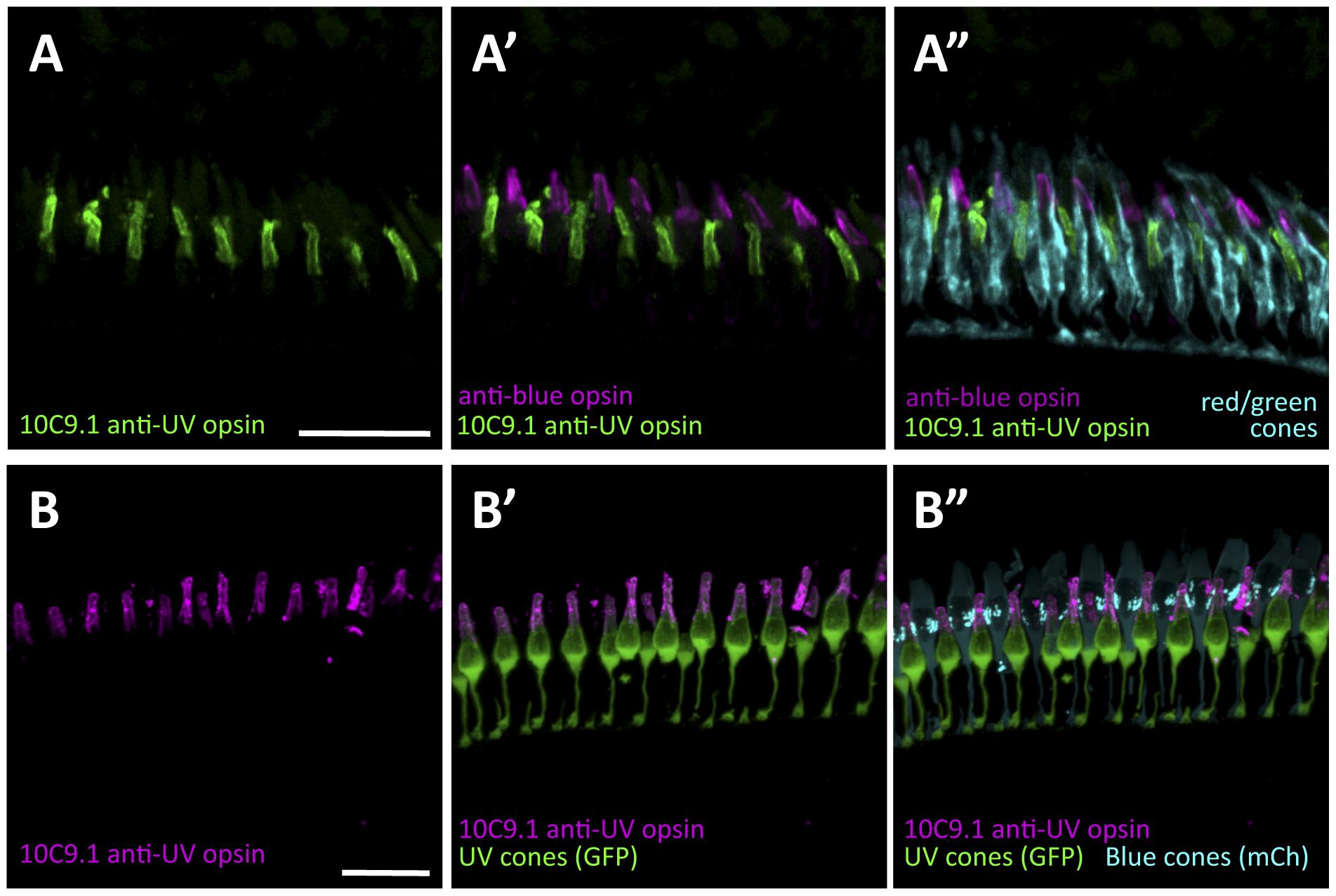
Fig. 5
A monoclonal antibody raised in rat (10C9.1) labels zebrafish UV cone outer segments, allowing all cone subtypes to be simultaneously labeled by immunohistochemistry.
Antibody 10C9.1 specifically labels the outer segments of a class of short single cones in the adult zebrafish retina (Figure S1 panel A). 10C9.1 specificity is supported (see Figure S1 panels B–D), including by a dramatic decrease in number of cells labeled when 10C9.1 is applied to retinas from zebrafish mutants (tbx2blor/lor) that have a paucity of UV cones. A. The population of single cones labeled by 10C9.1 is the UV cones, because established antibodies against the other single cone class, the blue cones, labels a distinct cone population (A2). 10C9.1 enables an unprecedented combination of antibodies raised in different species that simultaneously label and distinguish all cone photoreceptor subtypes (A′′). E. Further evidence that 10C9.1 labels UV cone outer segments comes from its co-localization with UV cones filled with green fluorescent protein (GFP), and its exclusion from blue cones filled with mCherry (mCh) in transgenic zebrafish (Tg(-5.5opn1sw1:EGFP)kj9;Tg(-3.5opn1sw2:m Cherry)ua3011). Panel B is available as Movie S1. Scale bars 30 μm.