
|
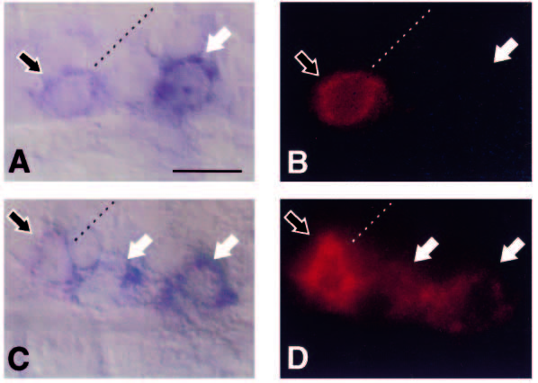
Fig. 6 Simultaneous detection of islet1 and islet2 RNA in primary motoneurons. (A and C) Double labels for islet1 RNA (magenta) and islet2 RNA (blue). (B and D) Corresponding fluorescent images showing low levels of islet1 RNA. Photos are from a 21-somite-stage embryo. Anterior is left and dorsal is up. Dashed lines represent approximate positions of somite borders. Scale bar equals 10 μm. (A,B) At somite level 14, only islet1 is expressed in a cell near the somite border (outlined arrow) and islet2, alone, is expressed in a midsegment cell (white arrow). (C,D) At somite level 17, islet1, and not islet2, RNA is detected in a cell near the somite border (outlined arrow) while both islet1 and islet2 RNAs are detected in two midsegment cells (white arrows).