
|
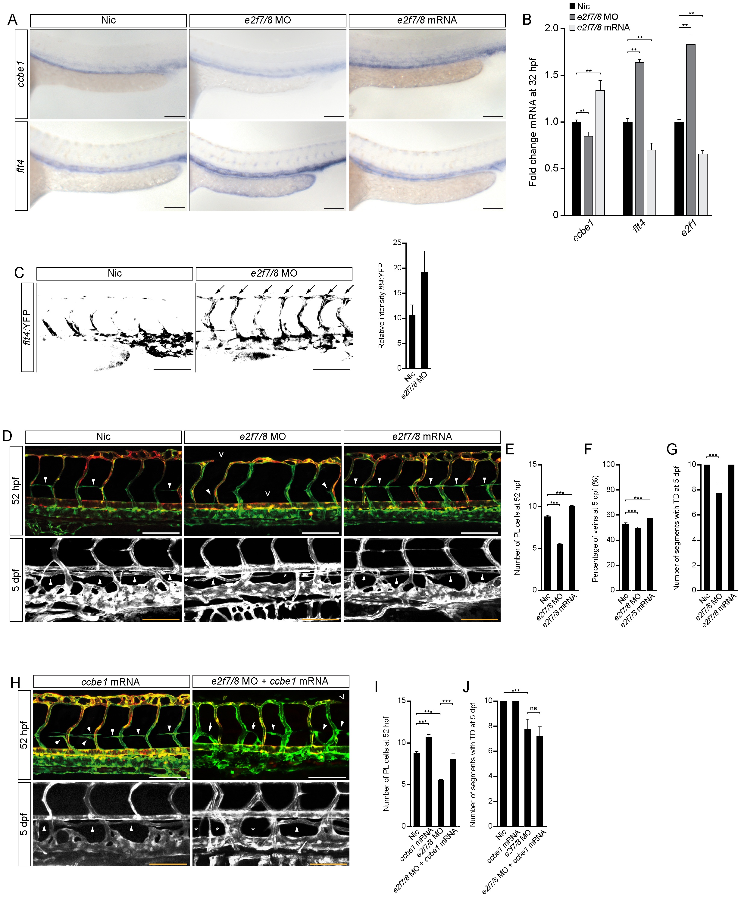
Fig. 2 Loss of E2f7/8 impaired venous sprouting and lymphangiogenesis.
A, In situ hybridisation and B, qPCR (** P<0.05; two independent experiments with n = 10 per condition and experiment) for flt4 and ccbe1 in zebrafish embryos 32 hpf, un-injected control (nic) or injected with e2f7/8 MOs or mRNA. C, Flt4:YFP transgene level of 36 hpf uninjected or e2f7/8 MOs injected embryos, lateral view (n = 30 per condition). D–G Lateral images and quantification of Tg(fli1a:gfp;flt1enh:rfp) embryos treated as indicated and imaged at 52 hpf or 5 dpf. H–J Lateral images and quantification of Tg(fli1a:gfp;flt1enh:rfp) embryos treated as indicated and imaged at 52 hpf or 5 dpf. Concentrations: e2f7/8 MOs (10 ng each); e2f7/8 mRNA (100 pg each); ccbe1 mRNA (100 pg). Open arrow heads indicate missing intersegmental vessels or dorsal longitudinal anastomotic vessel. Closed arrow heads indicate PLs (upper panel) or the TD (lower panel). Arrows depict PLs that have connected to ISVs. Stars indicate missing TD fragments. All scale bars are 100 μm. Data presented as the average (±s.e.m.) compared to the control condition in three independent experiments (*** P<0.001). At least n = 150 embryos per condition in three independent experiments were used for D–J.