
|
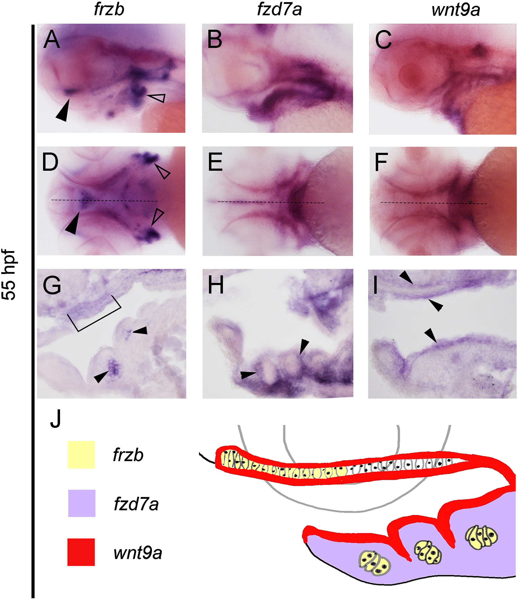
Fig. 1 Expression patterns of wnt9a, frzb, and fzd7a within pharyngeal structures. (A–K) Whole-mount in situ hybridization with embryos oriented with head towards the left in lateral view (A, B, C), ventral views (D, E, F) and sagittal sections (G, H, I). Midline sagittal section plane is indicated by dotted line in D–F. frzb transcripts localize to the chondrocytes located in the distal portion of the palate (arrow in A, bracket in G), lower jaw (arrow in G) and lateral ceratobrachials (open arrows in A and D). Expression of fzd7a is restricted to the neural crest tissue surrounding the chondrocytes (B, E, arrows in H). wnt9a is expressed in oropharyngeal ectoderm, and in the tissue above the palate (C, F, arrows in I). Diagram summary of the expression domains and their spatial relationship (J).
Reprinted from Developmental Biology, 381(2), Kamel, G., Hoyos, T., Rochard, L., Dougherty, M., Tse, W., Shubinets, V., Grimaldi, M., and Liao, E.C., Requirement for frzb and fzd7a in cranial neural crest convergence and extension mechanisms during zebrafish palate and jaw morphogenesis, 423-33, Copyright (2013) with permission from Elsevier. Full text @ Dev. Biol.