
|
Fig. 2
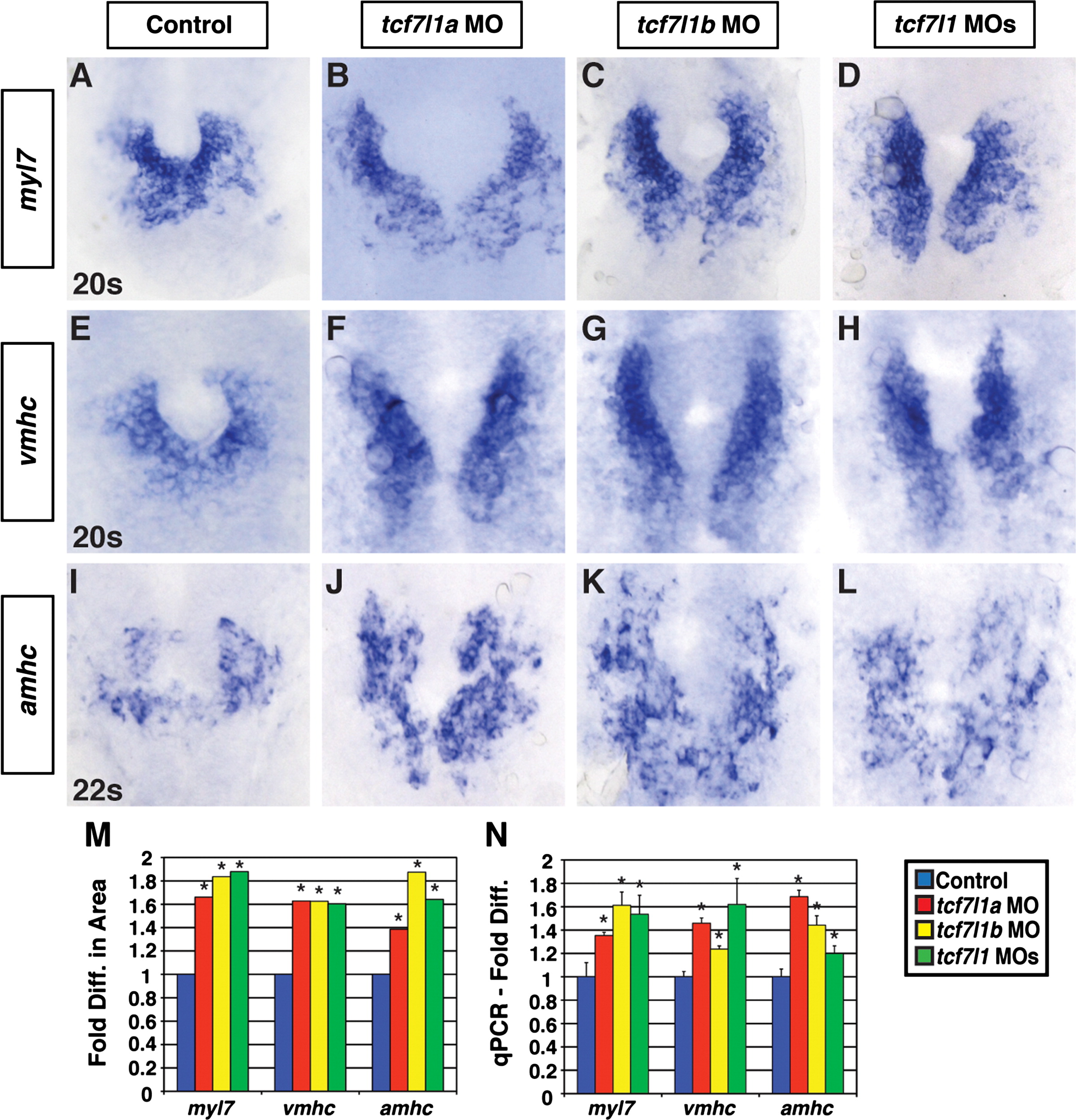
Tcf7l1a and Tcf7l1b restrict CM differentiation marker expression. ((A)–(D)) Myl7, ((E)–(H)) vmhc, and ((I)–(L)) amhc expression via ISH in control, Tcf7l1a, Tcf7l1b, and Tcf7l1 deficient embryos at the 20s and 22s stages. In contrast to CM counts at 48 hpf, similar increases in the CM differentiation markers were observed in the individual Tcf7l1a and Tcf7l1b depleted embryos compared to the Tcf7l1 depleted embryos. (M) Fold difference in the areas of cells expressing the CM differentiation marker genes. For analysis of myl7, control n=38, Tcf7l1a deficient n=25, Tcf7l1b deficient n=22, and Tcf7l1 deficient n=26. For analysis of vmhc, control n=32, Tcf7l1a deficient n=23, Tcf7l1b deficient n=23, and Tcf7l1 deficient n=28. For analysis of amhc, control n=37, Tcf7l1a deficient n=26, Tcf7l1b deficient n=35, and Tcf7l1 deficient n=22. (N) qPCR for CM differentiation marker gene expression at the 22s stage. For both the area measurements and qPCR, amhc displayed more experimental variability than myl7 and vmhc, but did not reveal an increase in differentiation over the individually depleted Tcf7l1a or Tcf7l1b embryos as found counting atrial cells at a later stage. Images are from flatmounted embryos. Views are dorsal with anterior up. Asterisk indicates a statistically significant difference compared to controls.
Reprinted from Developmental Biology, 380(2), Sorrell, M.R., Dohn, T.E., D'Aniello, E., and Waxman, J.S., Tcf7l1 proteins cell autonomously restrict cardiomyocyte and promote endothelial specification in zebrafish, 199-210, Copyright (2013) with permission from Elsevier. Full text @ Dev. Biol.