
|
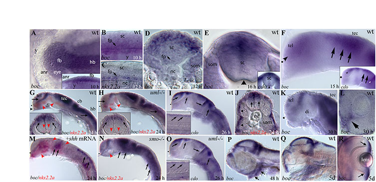
Fig. S1 boc and cdo expression during zebrafish development. (A) Dorsal view of the head, anterior left. Inset shows lateral view. At 10 hours postfertilization (hpf), boc is expressed in the presumptive forebrain, eye and central nervous system, with higher expression ventrally (inset). (B,C) Lateral views of the trunk, anterior left. (B) boc is first expressed in the ventral spinal cord at -10 hpf (arrow). (C) Expression of the Hh target gene nkx2.2a is first detected in the ventral CNS between 12 and 14 hpf (arrow). (D,E) Cross-sections through the trunk. (D) By 12 hpf boc is strongly expressed in the dorsal CNS, with weaker ventral expression remaining (arrow). boc is also expressed in the somites. (E) boc continues to be expressed in the dorsal spinal cord at 16 hpf. cdo is expressed in a similar dorsal domain (inset). Arrowhead marks the floor plate.(F-I) Lateral views of the head, eyes removed. (F) At 15 hpf, boc is regionally expressed in the telencephalon (arrowhead), dorsal midbrain (arrows) and hindbrain. cdo is expressed in a more restricted dorsal domain than boc (inset). (G) At 24 hpf, dorsal boc expression (arrows) is complementary to ventral nkx2.2a expression (red arrowheads) in the brain and spinal cord (inset, in cross-section). Low levels of boc expression can be detected in the region of the developing pituitary (black arrowhead). (H) In uml mutants identified by defects in nkx2.2a expression (red arrowheads), boc expression is slightly reduced in the dorsal brain and spinal cord (arrows). (I) cdo expression is similar to boc expression, with the cdo expression domain being more dorsally restricted in the brain (arrows) and spinal cord (inset, lateral view of trunk). (J) Cross-section through the trunk showing boc expression in the dorsal spinal cord (arrows) and in slow muscle fibers of the lateral somites (arrowheads). (K) At 30 hpf boc is expressed in the telencephalon, dorsal anterior diencephalon and tectum. (L) At 30 hpf boc is also expressed in the choroid fissure of the eye (arrow). (M) boc expression is dramatically downregulated in shh mRNA-injected embryos (arrows) whereas nkx2.2a expression is expanded dorsally (red arrowheads). (N) boc expression is expanded ventrally in the Hh pathway mutant smoothened (arrows) whereas nkx2.2a expression is absent (red arrowhead). (O) cdo expression is relatively unchanged in uml mutants compared with wild type (inset shows lateral view of trunk). (P) At 48 hpf boc is regionally expressed in the brain and in the developing branchial arches (arrows). (Q,R) At 5 days post fertilization (dpf) boc continues to be expressed in the dorsal CNS (Q) and in the lateral margin of the eye (R, arrows). Dots mark the optic recess in F-I and M-O. anr, anterior neural ridge; cb, cerebellum; di, diencephalon; fb, forebrain; fp, floor plate; hb, hindbrain; nc, notochord; sc, spinal cord; som, somite; tec, tectum; tel, telencephalon; y, yolk.