
|
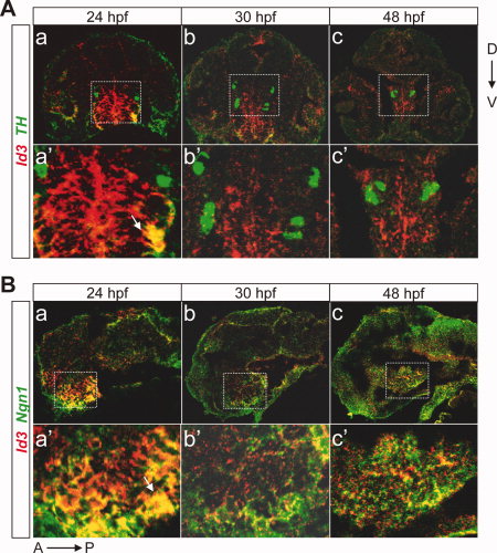
Fig. 4 Id3 is expressed in the diencephalon and partially colocalized with Ngn1 or TH. A: Double labeled fluorescent in situ hybridization in coronal cryosections of larvae at 24, 30, and 48 hours postfertilization (hpf) reveals that Id3 (red) and TH (green) are co-expressed in a subset of cells in the diencephalon. a′–c′: The magnified views of the stippled boxes in a–c. B: Double labeling of Id3 and Ngn1 in sagittal cryosections of larvae at 24, 30, and 48 hpf. a–c, sagittal views of the embryos after double fluorescent in situ hybridization of Id3 (red) and Ngn1 (green). a′–c′, the magnified views of the stippled boxes in a–c. Arrows indicate the cells with colocalized hybridization signals. TH, tyrosine hydroxylase; D, dorsal; V, ventral; A, anterior; P, posterior.