
|
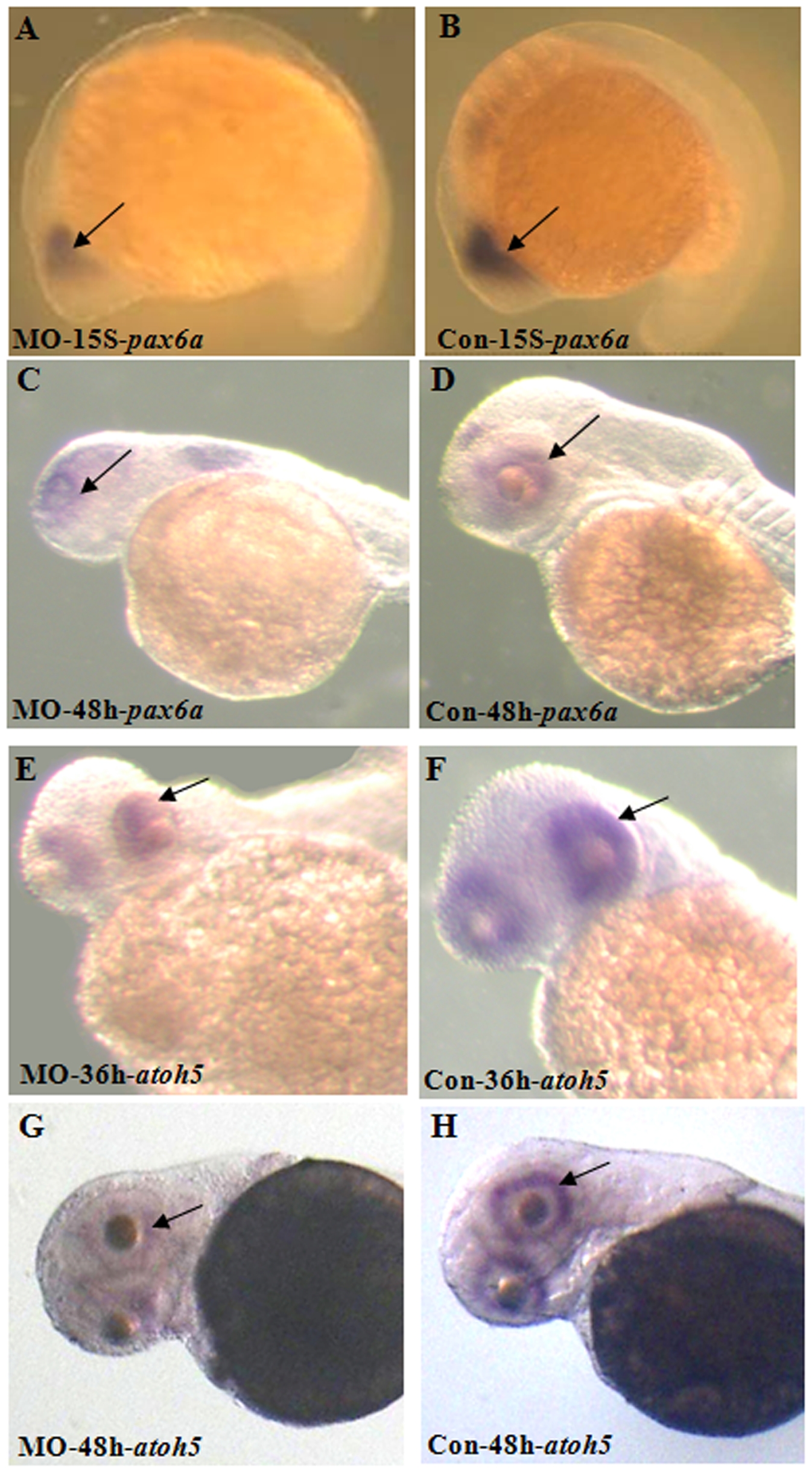
Fig. 10 Retinal development is disrupted in Atoh8 knockdown embryos.
(A–B) 15-Somite stage; (C–D,G–H) 48 hpf; (E–F) 36 hpf, (A,C,E,G) morphant and (B,D,F,H) control. Staining of pax6a showed that retinal region was remarkably reduced (arrow) in this MO-treated embryo. Atoh5 expression was present throughout most of the neural retinal at 36 hpf in the control embryo(E) while present only in dorsal and temporal retina in the MO-treated embryo(F). By 48 hpf, atoh5 localized throughout retina ganglion cells in the control(G) but present only in ventral retina in the MO-treated embryo(H). In addition, the signals of these two genes were much weaker than those of the control, indicating that knock-down of Atoh8 may have influenced retinal organization. In all panels, the dorsal side is up and the anterior is to the left.