
Image
|
Figure Caption
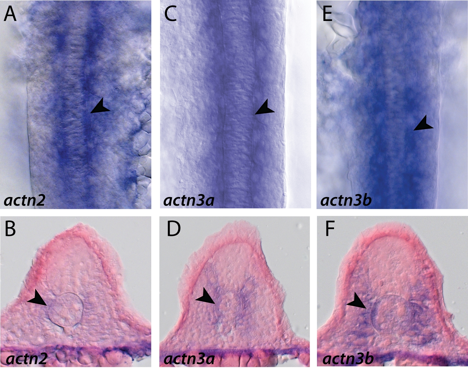
Fig. S3 Expression of the muscle actinins at 15 somites as shown by in situ hybridization. A, C, E: Dorsal views of 15 somite flatmounted embryos. B, D, F: Transverse sections taken at a level between somites 5 to 10. A-F: Expression of all three muscle actinins can be observed in the adaxial cells adjacent to the notocord (arrowheads). actn3a and actn3b are expressed more strongly and in a domain that has expanded slightly more dorsolaterally and ventrolaterally than actn2 at 15 somites (D,F).
Figure Data
Acknowledgments
This image is the copyrighted work of the attributed author or publisher, and
ZFIN has permission only to display this image to its users.
Additional permissions should be obtained from the applicable author or publisher of the image.
Full text @ Dev. Dyn.