
|
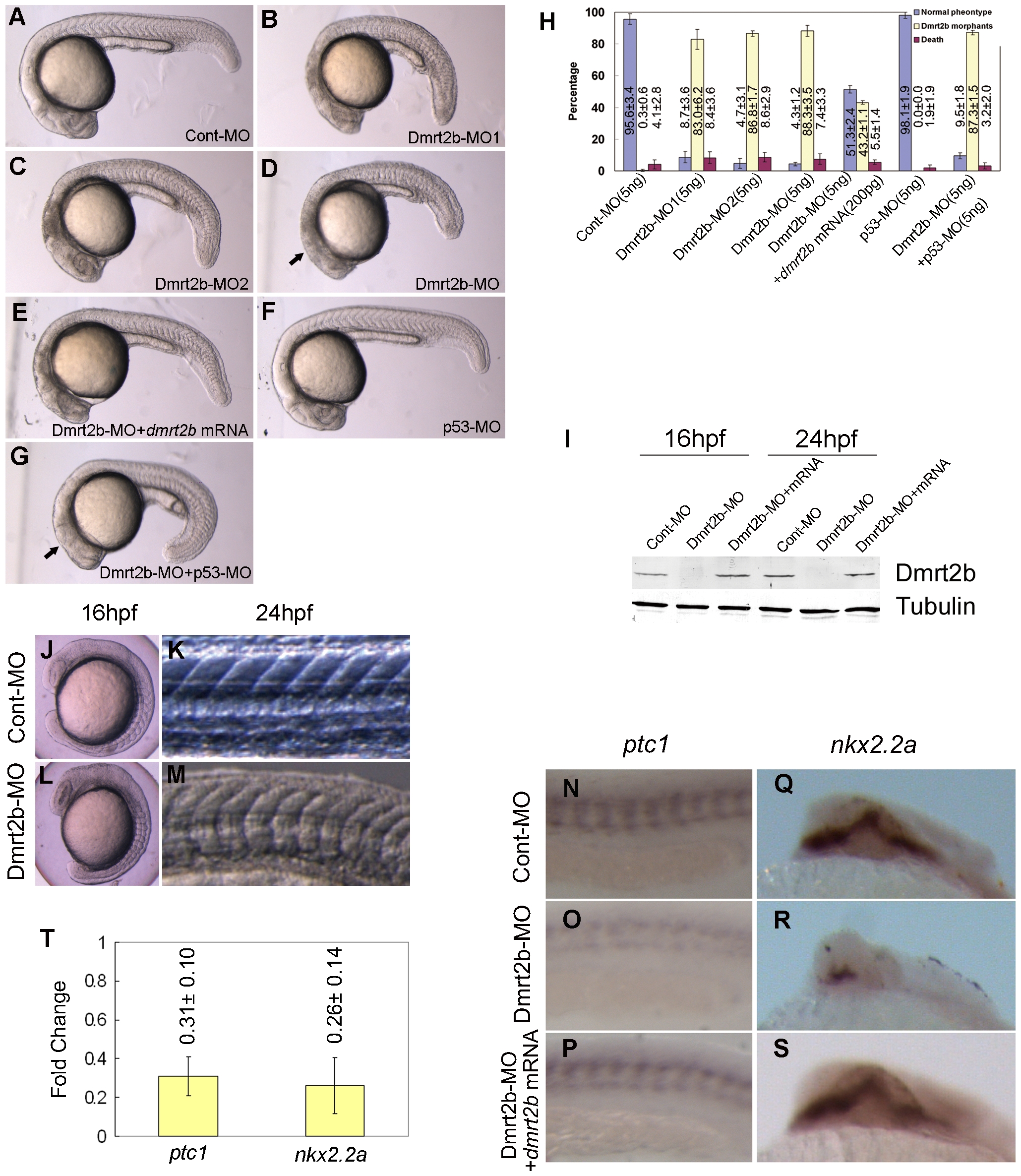
Fig. 2 Dmrt2b morphants display defects in somitogenesis and Hedgehog signaling.
(A–G) Morphology of 24 hpf embryos injected with Cont-MO (A), Dmrt2b-MO1 (B), Dmrt2b-MO2 (C), Dmrt2b-MO (D), Dmrt2b-MO+dmrt2b mRNA (E), p53-MO (F), Dmrt2b-MO+p53-MO (G). The arrows indicate the off-target cell death in Dmrt2b-MO morphant and the reduced cell death in the Dmrt2b-MO+p53-MO morphant. (H) The statistical data of three independent experiments on Dmrt2b knockdown, dmrt2b mRNA rescue and p53 MO co-injection. Results are represented as mean±SD of three separate experiments. (I) Western blot detection of Dmrt2b knockdown during embryogenesis. The protein extracts from embryos (16 hpf and 24 hpf) were analyzed by Western blot using the polyclonal anti-Dmrt2b antibody. A band of about 41 KD was not detected in Dmrt2b morphants. The picture represents typical result from three separate experiments. (J–M) Dmrt2b morphant exhibits U-shape somites. Morphology of embryos injected with Cont-MO display the typical ‘chevron’ shape (J, K). Morphology of embryos injected with Dmrt2b-MO display the U-shape (L, M). Whole-mount in situ hybridization of ptc1(N, O, P) (Anterior is left) and nkx2.2a(Q, R, S) (Anterior is top) in embryos injected with Cont-MO (N, Q) or Dmrt2b-MO (O, R) and embryos co-injected with Dmrt2b-MO with dmrt2b mRNA (P, S) at 24 hpf. (T) qPCR analysis of the expression changes of ptc1 and nkx2.2a in 24 hpf embryos injected with Cont-MO or Dmrt2b-MO. Results represent mean±SD of three separate experiments.