
|
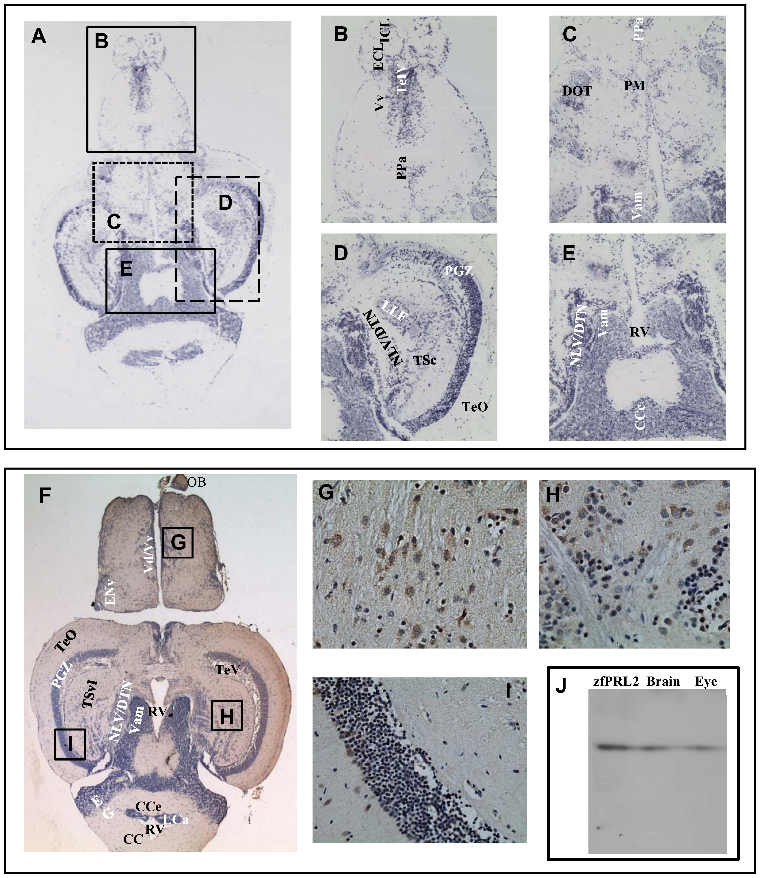
Fig. 5 Expression of PRL2 in zebrafish brain.
Horizontal sections of the zebrafish brain showing the expression of zfPRL2 after ISH and IHC. (A) An orientation photomicrograph showing the expression of zfPRL2 by ISH in a horizontal section through the zebrafish brain. (B–E) are photographs at higher magnification of the corresponding boxed areas shown in (A). (F) An orientation photomicrograph showing the expression of zfPRL2 by IHC in a horizontal section through the zebrafish brain. Brain regions with positive signals (brown dots) including dorsal nucleus of ventral telencephalic area/ventral nucleus of ventral telencephalic area (Vd/Vv), nucleus lateralis valvulae/dorsal tegmental nucleus (NLV/DTN) and periventricular gray zone of optic tectum (PGZ) are magnified in photomicrographs (G), (H) and (I) respectively. In both the ISH and IHC results, positive signals (dark blue dots) are observed in the following brain regions: corpus cerebella (CCe); dorsomedial optic tract (DOT); external cellular layer of olfactory bulb including mitral cells (ECL); internal cellular layer of olfactory bulb (ICL); lateral longitudinal fascicle (LLF); NLV/DTN; PGZ; magnocellular preoptic nucleus (PM); parvocellular preoptic nucleus, anterior part (PPa); rhombencephalic ventricle (RV); telencephalic ventricles (TeIV); tectum opticum (TeO); central nucleus of torus semicircularis (TSc); medial division of valvula cerebella (Vam); and ventral nucleus of ventral telencephalic area (Vv). Brain regions were identified on a topological atlas of the zebrafish brain [43]. Nuclei were counterstained with hematoxylin. A and F: x100; B–E: x200; G–I: x400. (J) Western blot showing expression of zfPRL2 in zebrafish brain and eye extracts.