
Image
|
Figure Caption
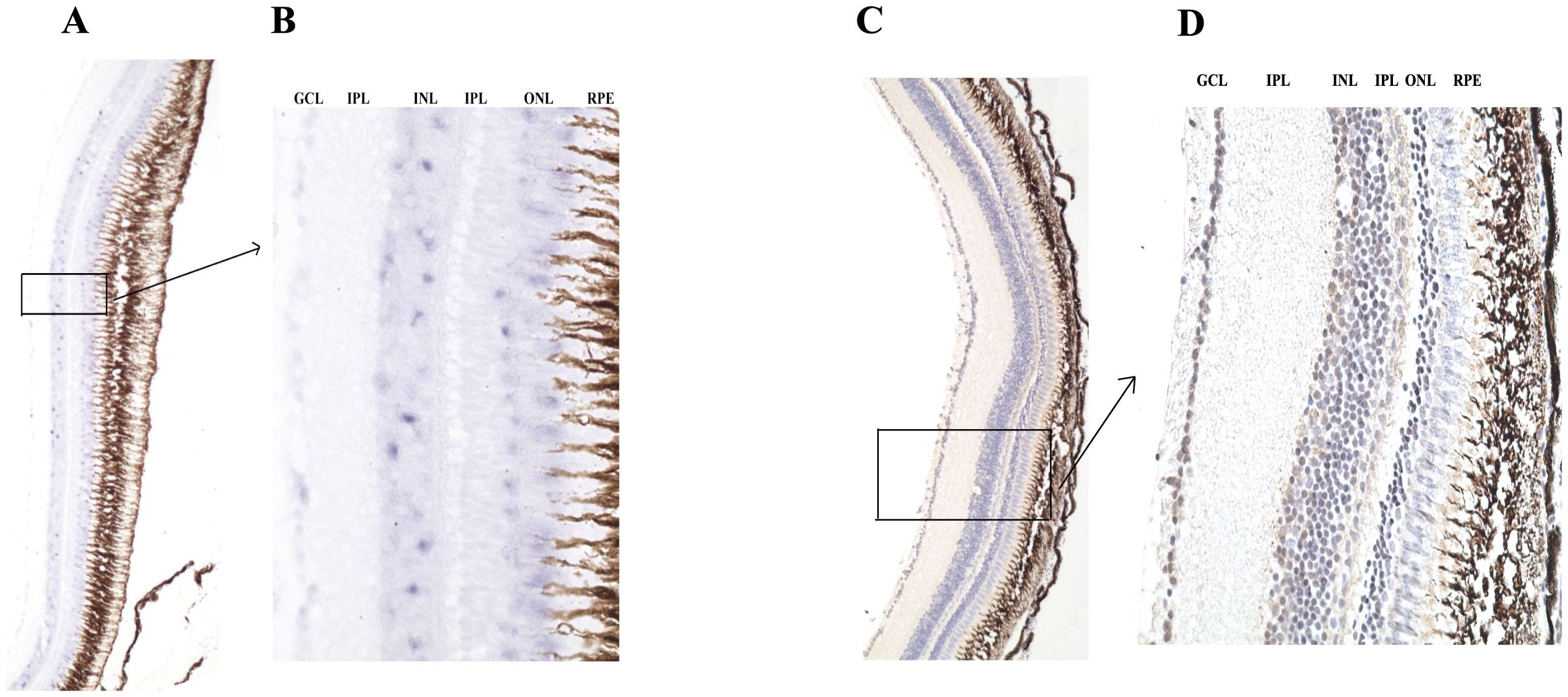
Fig. 4 Expression of PRL2 in zebrafish retina.
Transverse sections of the zebrafish retina showing expression of zfPRL2 after ISH and IHC. (A and B) ISH signals (gray blue dots) in the GCL, INL and ONL. (C and D) IHC signals (brown dots) in the GCL, INL and ONL. For IHC, nuclei were counterstained with hematoxylin. A and C: x100; B and D: x400. IPL: inner plexiform layer; OPL: outer plexiform layer; RPE: retinal pigmented epithelium.
Figure Data
Acknowledgments
This image is the copyrighted work of the attributed author or publisher, and
ZFIN has permission only to display this image to its users.
Additional permissions should be obtained from the applicable author or publisher of the image.
Full text @ PLoS One